Review
. 2011 Apr;3(4):385-428.
doi: 10.3390/nu3040385. Epub 2011 Mar 29.

Vitamin A in reproduction and development

Affiliations
Review

Vitamin A in reproduction and development

Margaret Clagett-Dame et al. Nutrients. 2011 Apr.

Abstract

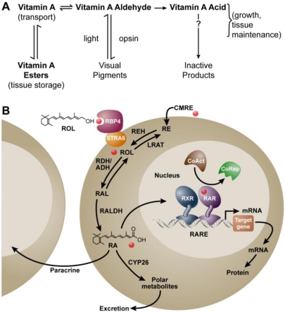
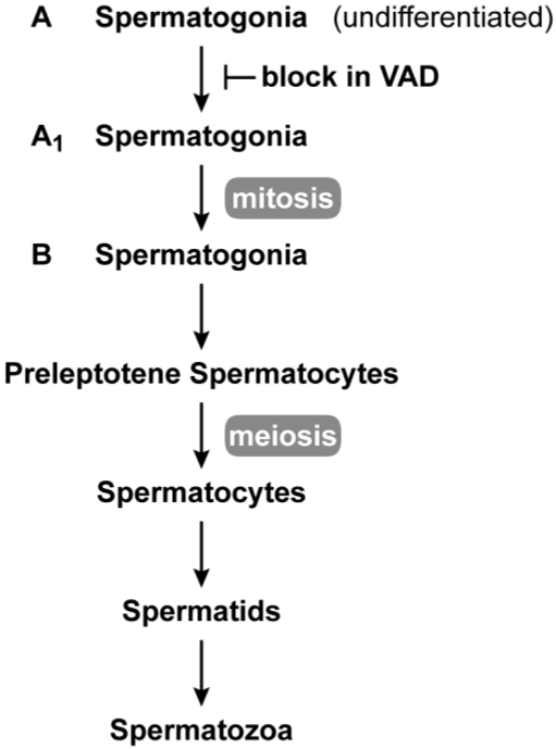
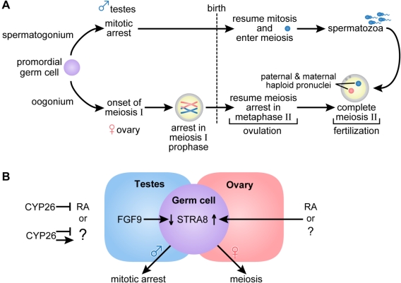
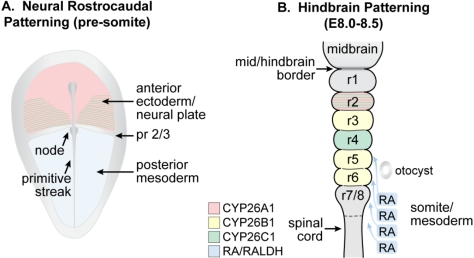
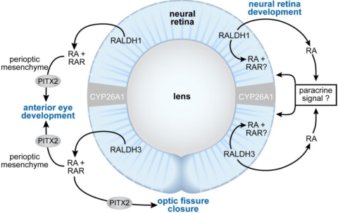
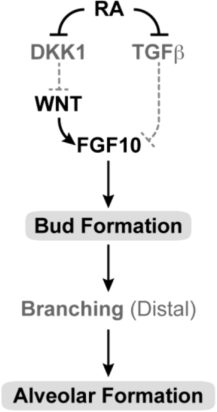
The requirement for vitamin A in reproduction was first recognized in the early 1900's, and its importance in the eyes of developing embryos was realized shortly after. A greater understanding of the large number of developmental processes that require vitamin A emerged first from nutritional deficiency studies in rat embryos, and later from genetic studies in mice. It is now generally believed that all-trans retinoic acid (RA) is the form of vitamin A that supports both male and female reproduction as well as embryonic development. This conclusion is based on the ability to reverse most reproductive and developmental blocks found in vitamin A deficiency induced either by nutritional or genetic means with RA, and the ability to recapitulate the majority of embryonic defects in retinoic acid receptor compound null mutants. The activity of the catabolic CYP26 enzymes in determining what tissues have access to RA has emerged as a key regulatory mechanism, and helps to explain why exogenous RA can rescue many vitamin A deficiency defects. In severely vitamin A-deficient (VAD) female rats, reproduction fails prior to implantation, whereas in VAD pregnant rats given small amounts of carotene or supported on limiting quantities of RA early in organogenesis, embryos form but show a collection of defects called the vitamin A deficiency syndrome or late vitamin A deficiency. Vitamin A is also essential for the maintenance of the male genital tract and spermatogenesis. Recent studies show that vitamin A participates in a signaling mechanism to initiate meiosis in the female gonad during embryogenesis, and in the male gonad postnatally. Both nutritional and genetic approaches are being used to elucidate the vitamin A-dependent pathways upon which these processes depend.

Keywords: retinoic acid; embryonic; vitamin A deficiency.


Figures

Figure 1
Figure 1
Metabolism of vitamin A (retinol) to all-trans retinoic acid (RA), and the mechanism of RA action. (A) Metabolic scheme proposed by Dowling and Wald in 1960 [11]; (B) Mechanism ca. 2011. Vitamin A (retinol, ROL) circulates bound to the plasma retinol-binding protein (RBP4) and transthyretin (not shown). RBP4 binds to the membrane receptor STRA6 to facilitate the cellular uptake of retinol in some cells. Vitamin A circulating as part of a chylomicron remnant (CMRE) can also serve as a source of vitamin A for the cell. Note that cellular retinol and RA binding proteins have been omitted for simplicity. Retinol is either esterified by lecithin:retinol acyltransferase (LRAT) and stored, or is oxidized reversibly to retinaldehyde (RAL) by retinol dehydrogenases (RDH/ADH), and further oxidized in irreversible fashion to RA by retinaldehyde dehydrogenase (RALDH 1, 2, or 3). In the nucleus, the RAR/RXR complex is bound to a specific sequence of DNA called the retinoic acid response element (RARE). Binding of RA to the RAR leads to release of the corepressor complex (CoRep) and association with coactivator proteins (CoAct), followed by altered transcription of downstream target genes and ultimately changes in cellular function. RA also undergoes further oxidation by the cytochrome P450 (CYP) 26 family to more polar metabolites. The lipophilic molecule, RA, can act within the same cell in which it is synthesized (autocrine) or can diffuse through the cell membrane to act in nearby cells (paracrine). Abbreviations: ADH, alcohol dehydrogenase; RDH, retinol dehydrogenase; REH, retinyl ester hydrolase; RE, retinyl ester.
Figure 2
Figure 2
Spermatogenesis in the adult. Spermatogenesis takes place in the seminiferous epithelium of testis tubules from puberty through adulthood. Undifferentiated (A-type) spermatogonia at the base of the seminiferous epithelium divide mitotically until they enter the differentiation pathway to become A1 spermatogonia. A1 spermatogonia undergo division to A1-A4 and finally B spermatogonia. B spermatogonia divide to produce preleptotene (primary) spermatocytes that migrate away from the base of the seminiferous tubule to undergo meiosis. The first meiotic division produces secondary spermatocytes, and after the second meiotic division, spermatids (haploid cells) begin the differentiation process (spermiogenesis) to spermatozoa. In vitamin A deficiency, the transition from A to A1 spermatogonia is blocked [42,43,44].
Figure 3
Figure 3
(A) Germ cell development and gametogenesis. Primordial germ cells colonize the gonad in both male and female embryos. The first morphological marker of sex-specific germ cell development is seen in the female embryo when the oogonia enter meiosis. Primary oocytes proceed through the leptotene, zygotene and pachytene stages of meiotic prophase before birth, when they arrest in diplotene of meiosis I. At ovulation, meiosis I is completed, and the secondary oocytes enter meiosis II and arrest again in metaphase. Meiosis II is completed after fertilization. In the male embryo, germ cells are committed to the spermatogenic program but arrest in G0/G1, and do not complete mitosis and enter meiosis until after they are born. Primary spermatocytes entering meiosis I are seen during the first week of life; secondary spermatocytes complete meiosis II forming spermatids and functional gametes called spermatozoa or sperm. In the male, waves of meiosis continue throughout life. (B) In the female embryo access to RA or alternatively, another factor indicated as (?), promotes entry into meiosis whereas embryonic male germ cells are maintained in a pluripotent state. Either RA or another factor acts in the embryonic female germ cell to increase Stra8, essential for entry into meiosis. In the ovary, Fgf9 levels are low. In the male embryo, entry into meiosis is prevented by the action of CYP26B1; high levels of Fgf9 in the testes antagonize Stra8 expression and maintain germ cells in a pluripotent state. (Adapted from [77,78]).
Figure 4
Figure 4
Schematic showing the location of RA and Cyp26 expression in a presomitic mouse embryo undergoing (A) patterning of the neural plate and (B) later during the course of hindbrain patterning. (A) RA generated by RALDH2 in the posterior mesoderm forms an early anterior boundary of activity in the neural plate at presumptive rhombomere (pr) 2/pr3, whereas Cyp26A1 and Cyp26C1 are expressed rostral to the pr2 border; (B) By E8.0 to E8.5, RA is being expressed by the somites and anterior presomitic mesoderm, and acts on the overlying hindbrain and spinal cord. The activity of the CYP26 enzymes regulate access of the neuroepithelium to RA (Adapted from [139]).
Figure 5
Figure 5
Schematic showing the proposed sites of RA function during eye morphogenesis (left) and differentiation (right). At early stages of eye development, RA generated by RALDH1 and RALDH3 acts as a paracrine signal binding to RARs located in the perioptic mesenchyme to support anterior eye segment development and closure of the optic fissure. Pitx2 is a RA/RAR-regulated transcription factor that is required both for anterior eye segment morphogenesis, as well as closure of the optic fissure. At later stages of development, RA promotes differentiation of the neural retina. The mechanism is unclear, but could involve either a paracrine effect of RA outside of the neural retina, or a direct effect on the cells within the retina itself.
Figure 6
Figure 6
Schematic showing the proposed sites of RA action in lung development. In the developing embryo, RA is needed for primary bud formation (grey oval), but signaling is down-regulated during the time that lung differentiation occurs. A later role for RA in alveolar formation (grey oval) is also proposed. During induction of the lung buds, RA regulates mesodermal Fgf10 levels by negatively regulating Tgfβ and enabling induction of the Wnt pathway by repression of the Dickkopf homolog 1 (Dkk1) known to antagonize Wnt ligand-receptor binding. RA may also influence the response of the foregut endoderm (origin of lung progenitors) to Fgf10. (Adapted from [262]).

Similar articles

Cited by

References

    1. McCollum E.V., Davis M. The necessity of certain lipins in the diet during growth. J. Biol. Chem. 1913;15:167–175. - PubMed
    1. Wolbach S.B., Howe P.R. Tissue changes following deprivation of fat-soluble A vitamin. J. Exp. Med. 1925;42:753–777. - PMC - PubMed
    1. Evans H.M. The effects of inadequate vitamin A on the sexual physiology of the female. J. Biol. Chem. 1928;77:651–654.
    1. Carpenter K.J., Harper A.E., Olson R.E. Experiments That Changed Nutritional Thinking. J. Nutr. 1997;127:1017S–1053S. - PubMed
    1. Moore T. Vitamin A and carotene: The absence of the liver oil vitamin A from carotene. VI. The conversion of carotene to vitamin A in vivo. Biochem. J. 1930;24:692–702. - PMC - PubMed

Publication types