. 2011 Dec;44(4):295-303.
doi: 10.5115/acb.2011.44.4.295. Epub 2011 Dec 30.

Different expressions of AQP1, AQP4, eNOS, and VEGF proteins in ischemic versus non-ischemic cerebropathy in rats: potential roles of AQP1 and eNOS in hydrocephalic and vasogenic edema formation

Affiliations

Different expressions of AQP1, AQP4, eNOS, and VEGF proteins in ischemic versus non-ischemic cerebropathy in rats: potential roles of AQP1 and eNOS in hydrocephalic and vasogenic edema formation

Jaehyun Kim et al. Anat Cell Biol. 2011 Dec.

Abstract

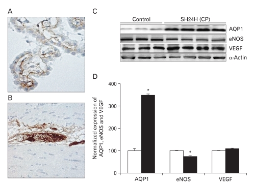
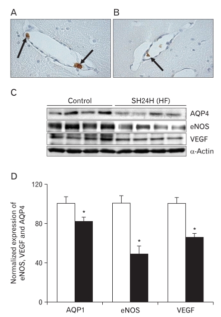
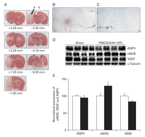
In this study, expressions of aquaporin (AQP) 1, AQP4, endothelial nitric oxide synthase (eNOS), and vascular endothelial growth factor in blood-cerebrospinal fluid (CSF) barrier and blood-brain barrier (BBB) are examined in rat choroid plexus and peri-infarcted hippocampal formation (HF) following systemic hyponatremia (SH) and permanent middle cerebral artery occlusion (pMCAO). These events are thought to cause the development of hydrocephalic and vasogenic edemas. The importance of CSF overproduction and intact blood-CSF barrier during hydrocephalic edema formation is demonstrated by the high expression of AQP1 (329.86±10.2%, n=4 , P<0.01) and trapped plasma immunoglobulin G (IgG) in choroid plexus epithelium after 24 hours of SH. However, the increased eNOS expression in peri-infarcted HF (130±3%, n=4, P<0.01) and extravasation of plasma IgG into the extravascular compartment after 24 hours of pMCAO suggest that increased microvascular permeability, probably due to elevated levels of nitric oxide, leads to development of vasogenic brain edema via BBB breakdown. Based on these findings, the authors suggest that modulation of different protein expression, dependent on the type of brain edema, is required for primary (pMCAO) and secondary (SH) brain injuries to attenuate brain edema and neuronal degeneration.

Keywords: Aquaporin 1; Blood-CSF barrier; Blood-brain barrier; Brain edema; eNOS.


Figures

Fig. 1
Fig. 1
Extravasation of plasma immuno globulin G (IgG) and the expressions of aquaporin 1 (AQP1), endothelial nitric oxide synthase (eNOS), and vascular endothelial growth factor (VEGF) in choroid plexus after 24 h of systemic hyponatremia (SH). (A, B) No plasma IgG extravasation is observed in the choroid plexus of third and lateral ventricles after 24 h of SH (×20). (C) Immunoblots are reacted with anti-AQP1, -eNOS, and -VEGF antibodies to reveal 28, 130, and 49 kDa products. α-Actin is used as an internal control. (D) Densitometric analysis reveals that SH significantly increases AQP1 expression in choroid plexus (329.86±10.2%, n=4, *P<0.01). In contrast, the expression of eNOS at 24 h after SH is significantly lower than that of control rats (75±3%, n=4, *P<0.01).
Fig. 2
Fig. 2
Extravasation of plasma immunoglobulin G (IgG) and the expressions of aquaporin4 (AQP4), endothelial nitric oxide synthase (eNOS), and vascular endothelial growth factor (VEGF) in the hippocampal formation (HF) after 24 h of systemic hyponatremia (SH). (A, B) Plasma IgG (arrows) is trapped solely in the luminal side of capillaries and/or venules in HF after 24 h of SH (×40). (C) Immunoblot with affinity purified anti-AQP4, -eNOS, and -VEGF antibodies, revealing 32, 130, and 49 kDa products. α-Actin is used as an internal control. (D) Densitometric analysis reveals that SH significantly decrease AQP4, eNOS, and VEGF expressions in HF compared with those of control rats (AQP4, 82±4%; eNOS, 49±8%; VEGF, 66±4%, n=4, *P<0.01).
Fig. 3
Fig. 3
Extravasation of plasma immunoglobulin G (IgG) and the expressions of aquaporin4 (AQP4), endothelial nitric oxide synthase (eNOS), and vascular endothelial growth factor (VEGF) in peri-infarcted hippocampal formation (HF) after 24 h of permanent middle cerebral artery occlusion (pMCAO). (A) 2,3,5-Triphenyltetrazolium chloride staining of brain slices from bregma +4.20 to -8.30 mm. Peri-infarcted HF (b, marked with a white arrow) represented by the red zone near the infarction zone (a, marked with black arrow) in the ipsilateral hemisphere over a series of brain sections. (B, C) Extensive positive staining of extravasated plasma IgG is seen at the ipsilateral peri-infarcted HF. However, no prominent positive profiles are identified in contralateral HF (×4). (D) This immunoblot is reacted with affinity purified anti-AQP4, -eNOS and -VEGF antibodies to reveal 32, 130, and 49 kDa products. β-Tubulin is used as an internal control. (E) Densitometric analysis reveals that pMCAO significantly increases eNOS (130±3%, n=4, *P<0.01) in peri-infarcted HF as compared with sham-operated controls. However, the expressions of AQP4 and VEGF are not changed after pMCAO.

References

    1. Kimelberg HK. Current concepts of brain edema. Review of laboratory investigations. J Neurosurg. 1995;83:1051–1059. - PubMed
    1. Blei AT. Pathophysiology of brain edema in fulminant hepatic failure, revisited. Metab Brain Dis. 2001;16:85–94. - PubMed
    1. Jung YW, Choi IJ, Kwon TH. Altered expression of sodium transporters in ischemic penumbra after focal cerebral ischemia in rats. Neurosci Res. 2007;59:152–159. - PubMed
    1. Klatzo I. Evolution of brain edema concepts. Acta Neurochir Suppl (Wien) 1994;60:3–6. - PubMed
    1. Papadopoulos MC, Manley GT, Krishna S, Verkman AS. Aquaporin-4 facilitates reabsorption of excess fluid in vasogenic brain edema. FASEB J. 2004;18:1291–1293. - PubMed