. 2012 Jan 1;3(1):37-47.
doi: 10.1364/BOE.3.000037. Epub 2011 Dec 5.

Extending vaterite microviscometry to ex vivo blood vessels by serial calibration

Affiliations

Extending vaterite microviscometry to ex vivo blood vessels by serial calibration

Samir G Shreim et al. Biomed Opt Express. .

Abstract

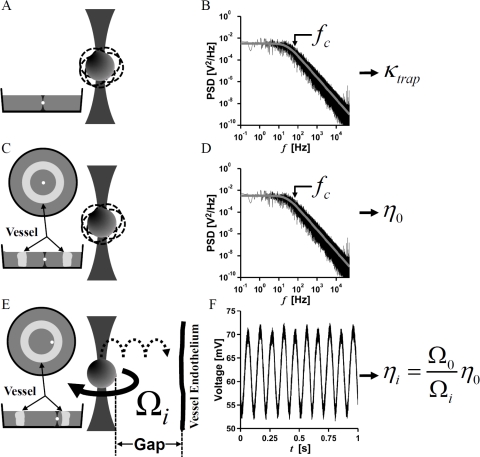
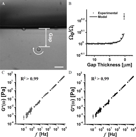
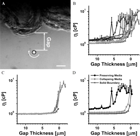
The endothelial glycocalyx layer is a ~2 µm thick glycosaminoglycan rich pericellular matrix expressed on the luminal surface of vascular endothelial cells, which has implications in vessel mechanics and mechanotransduction. Despite its role in vascular physiology, no direct measurement has of yet been made of vessel glycocalyx material properties. Vaterite microviscometry is a laser tweezers based microrheological method, which has been previously utilized to measure the viscosity of linear and complex fluids under flow. This form of microrheology has until now relied on complete recollection of the forward scattered light. Here we present a novel method to extend vaterite microviscometry to relatively thick samples. We validate our method and its assumptions and measure the apparent viscosity as a function of distance from the vascular endothelium. We observe a differential response in conditions designed to preserve the EGL in comparison to those designed to collapse it.

Keywords: (140.7010) Laser trapping; (160.1435) Biomaterials; (170.4520) Optical confinement and manipulation.


Figures

Fig. 1
Fig. 1
Schematic of experimental approach. Laser trap stiffness, κtrap, is first measured by trapping a particle of known diameter in water (A), and then determining the corner frequency from a Lorentzian fit to the power spectral density of particle fluctuations (B). κtrap is related to the corner frequency by Eq. (2). The procedure is repeated in the center of an excised vessel slice (C), where the corner frequency of the power spectral density (D) is used to determine the viscosity,η0of the culture media by Eq. (2). The apparent viscosity, ηias a function of gap thickness is determined by optically measuring the rotation rate of a vaterite particle rotating under constant optical torque at various distances from the vessel wall (E). For each gap thickness, ηi is calculated by measuring vaterite particle rotation rate and Eq. (5) (F).
Fig. 2
Fig. 2
Validating VMV near a glass boundary and linearity of our culture media. (A) A 4.5 µm vaterite microsphere rotating at 6.4 Hz in water at room temperature near a solid boundary comprising a glass wall. The gap thickness is measured as shown. Scale bar is 10 µm (B) Experimental data and fit of relative rotation rates as a function of gap thickness. The residual of fit is 0.0791, mostly due the point closest to the boundary. Note the fine scale on the vertical axis. (C) PMR measurement in EGL collapsing media. (D) PMR measurement in EGL preserving media. PMR confirms linearity of the culture media across observed frequencies.
Fig. 3
Fig. 3
VMV of the EGL in an excised porcine femoral artery. (A) A 4.7 µm vaterite microsphere rotating at 6.0 Hz in EGL preserving media near the endothelium of an excised vessel slice. The gap thickness is measured as shown. Scale bar is 10 µm. (B) Apparent viscosity, ηi measured in an excised vessel slice cultured in EGL preserving media (C) Apparent viscosity, ηi measured in an excised vessel slice cultured in EGL collapsing media. (D) Representative data for vessel experiments in EGL preserving and collapsing media, as well as the solid glass boundary experiment in water highlight role of the EGL in modulating local apparent viscosity.

Similar articles

Cited by

References

    1. Davies P. F., “Flow-mediated endothelial mechanotransduction,” Physiol. Rev. 75(3), 519–560 (1995). - PMC - PubMed
    1. Chien S., “Mechanotransduction and endothelial cell homeostasis: the wisdom of the cell,” Am. J. Physiol. Heart Circ. Physiol. 292(3), H1209–H1224 (2007).10.1152/ajpheart.01047.2006 - DOI - PubMed
    1. Reitsma S., Slaaf D. W., Vink H., van Zandvoort M. A. M. J., oude Egbrink M. G. A., “The endothelial glycocalyx: composition, functions, and visualization,” Pflugers Arch. 454(3), 345–359 (2007).10.1007/s00424-007-0212-8 - DOI - PMC - PubMed
    1. Weinbaum S., Tarbell J. M., Damiano E. R., “The structure and function of the endothelial glycocalyx layer,” Annu. Rev. Biomed. Eng. 9(1), 121–167 (2007).10.1146/annurev.bioeng.9.060906.151959 - DOI - PubMed
    1. Vink H., Duling B. R., “Identification of distinct luminal domains for macromolecules, erythrocytes, and leukocytes within mammalian capillaries,” Circ. Res. 79(3), 581–589 (1996). - PubMed