

doi: 10.1364/BOE.3.000153.
Epub 2011 Dec 16.
Measurement of the traction force of biological cells by digital holography
Affiliations
- PMID: 22254175
- PMCID: PMC3255332
- DOI: 10.1364/BOE.3.000153
Item in Clipboard
Measurement of the traction force of biological cells by digital holography
Biomed Opt Express.
.
Abstract
The traction force produced by biological cells has been visualized as distortions in flexible substrata. We have utilized quantitative phase microscopy by digital holography (DH-QPM) to study the wrinkling of a silicone rubber film by motile fibroblasts. Surface deformation and the cellular traction force have been measured from phase profiles in a direct and straightforward manner. DH-QPM is shown to provide highly efficient and versatile means for quantitatively analyzing cellular motility.
Keywords: (090.1995) Digital holography; (170.0180) Microscopy; (170.3880) Medical and biological imaging.
2011 Optical Society of America
Figures
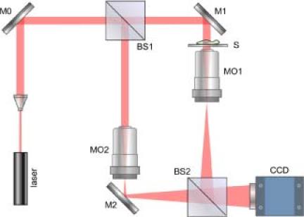
DHM setup. M’s: mirrors; BS’s: beam splitters; MO’s: microscope objectives; S: sample object
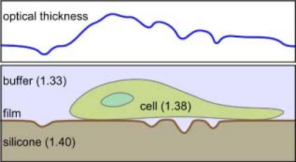
Schematic of the cell-substrate sample (lower) and the corresponding optical thickness profile (upper).
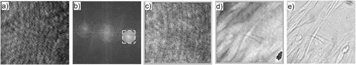
DHM analysis of fibroblasts wrinkling the silicone rubber film. The field of view is 190 × 176 μm2 with 800 × 742 pixels. a) Hologram; b) Angular spectrum; c) Amplitude image; d) Quantitative phase image; e) Bright field image.
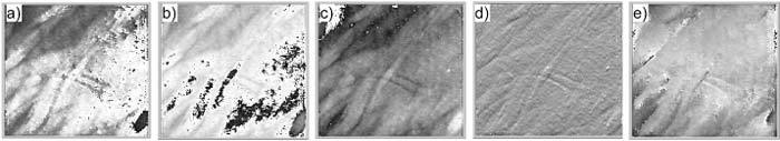
Multimode imaging from a single hologram. The field of view is 190 × 176 μm2 with 800 × 742 pixels. a) dark field; b) Zernike+; c) Zernike–; d) DIC; e) spiral DIC.
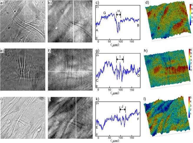
Examples of cells wrinkling a silicone rubber film. The field of view was 190 × 176 μm2 with 800 × 742 pixels. a), e) and i) Bright field images; b), f) and j) Quantitative phase images; c), g) and k) Cross-sections of phase profiles along highlighted lines AB in b), CD in f) and EF in j); d), h) and l) Pseudo-color 3-D rendering of phase images b), f) and j).
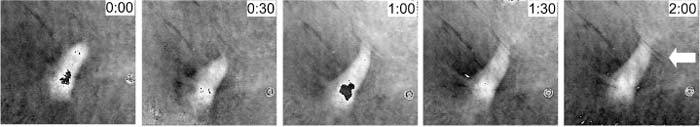
An excerpt of several frames from phase movie recordings of cells wrinkling a silicone rubber film (Media 1 ). The field of view was 190 × 176 μm2 with 800 × 742 pixels. Time interval of two contiguous images above was around 30 min.
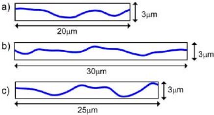
Phase profiles scaled as physical thickness and plotted in proportion to horizontal distance. a) Wrinkled area H from Fig. 5c). b) Wrinkled area I from Fig. 5g). c) Wrinkled area J from Fig. 3k). In each case, the average of 10 adjacent profiles is presented.
References
LinkOut - more resources
Full Text Sources