. 2011:2011:1672-5.
doi: 10.1109/IEMBS.2011.6090481.

Image-based estimation of ventricular fiber orientations for patient-specific simulations

Affiliations

Image-based estimation of ventricular fiber orientations for patient-specific simulations

Fijoy Vadakkumpadan et al. Annu Int Conf IEEE Eng Med Biol Soc. 2011.

Abstract

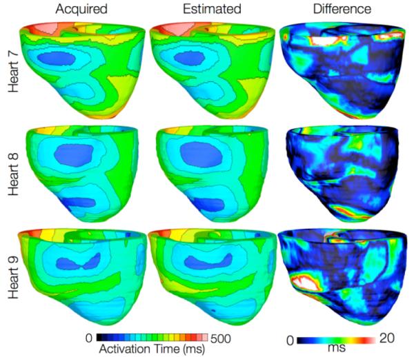
Patient-specific simulation of heart (dys)function aimed at personalizing cardiac therapy are hampered by the absence of in vivo imaging technology for clinically acquiring myocardial fiber orientations. In this research, we develop a methodology to predict ventricular fiber orientations of a patient heart, given the geometry of the heart and an atlas. We test the methodology by comparing the estimated fiber orientations with measured ones, and by quantifying the effect of the estimation error on outcomes of electrophysiological simulations, in normal and failing canine hearts. The new insights obtained from the project will pave the way for the development of patient-specific models of the heart that can aid physicians in personalized diagnosis and decisions regarding electrophysiological interventions.


Figures

Fig. 1
Fig. 1
Geometry and fiber orientations of the atlas ventricles. (A) The epicardial (red) and endocardial (green and magenta) splines, and corresponding landmarks (yellow) overlaid on an examples slice of the atlas image. (B) The atlas ventricles in 3D. (C) The atlas fiber orientations.
Fig. 2
Fig. 2
Application of the fiber orientation estimation methodology to an example patient heart image. (A) The epicardial (red) and endocardial (green and magenta) splines, and corresponding landmarks (yellow) overlaid on an image slice. (B) patient ventricles in 3D. (C) Superimposition of ventricles of atlas (magenta, see Fig. 1B) and patient. (D) Patient ventricles and the affine transformed atlas ventricles. (E) Patient ventricles and LDDMM-transformed atlas ventricles. (F) Estimated patient ventricular fiber orientations.
Fig. 3
Fig. 3
Validation of the fiber orientation estimation methodology by comparing estimated fiber orientations with DTMR-derived orientations. (A) Distribution of mean estimation error in normal hearts. (B) Distribution of mean estimation error in failing hearts.
Fig. 4
Fig. 4
Results from one beat of simulations of sinus rhythm in normal canine hearts. (A) Simulated activation map with acquired fiber orientations (model 1). (B) Absolute difference between simulated activation map with acquired fiber orientations and that with estimated fiber orientations, averaged over the five estimates. (C) Simulated activation maps with estimated fiber orientations (models 2-6)
Fig. 5
Fig. 5
Results from simulations of one beat of sinus rhythm in failing heart models. In the first column, rows 1-3 show simulated activation maps with models 7-9, respectively. In the second column, rows 1-3 display results of simulations with models 10-12, respectively. Rows 1-3 in the third column illustrate the absolute difference between the activation maps shown in the first and second columns of the corresponding row.

References

    1. Trayanova N. Whole heart modeling: Applications to cardiac electrophysiology and electromechanics. Circulation Research. 2011;108:113–128. - PMC - PubMed
    1. Sosnovik DE, Wang R, Dai G, Reese TG, Wedeen VJ. Diffusion mr tractography of the heart. Journal of Cardiovascular Magnetic Resonance. 2009;11:47–61. - PMC - PubMed
    1. Vadakkumpadan F, Arevalo H, Prassl AJ, Chen J, Kickinger F, et al. Image-based models of cardiac structure in health and disease. Wiley Interdisciplinary Reviews: Systems Biology and Medicine. 2010;2:489–506. - PMC - PubMed
    1. Beg MF, Miller MI, Trouve A. Computing large deformation metric mappings via geodesic flows of diffeomorphisms. International Journal of Computer Vision. 2005;61:139–157.
    1. Alexander DC, Pierpaoli C, Basser PJ, Gee JC. Spatial transformations of diffusion tensor magnetic resonance images. IEEE Transactions on Medical Imaging. 2001;20:1131–1139. - PubMed

Publication types

LinkOut - more resources