

Electrocortical source imaging of intracranial EEG data in epilepsy
- PMID: 22255194
- PMCID: PMC4135517
- DOI: 10.1109/IEMBS.2011.6090971
Electrocortical source imaging of intracranial EEG data in epilepsy
Abstract
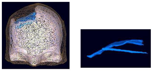
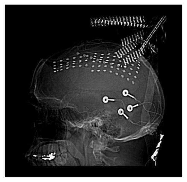
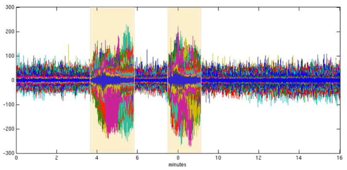
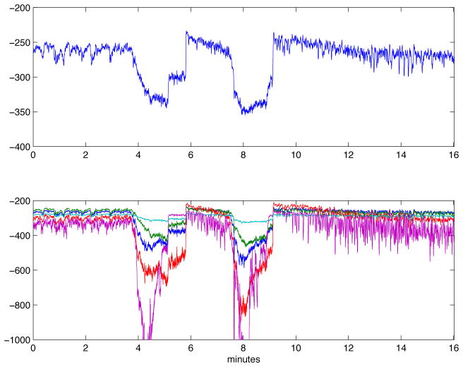
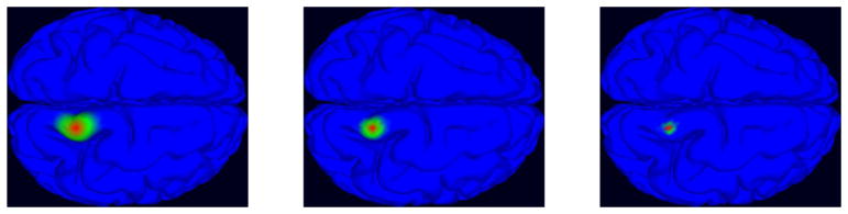
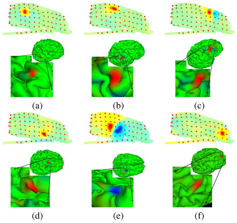
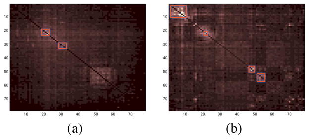
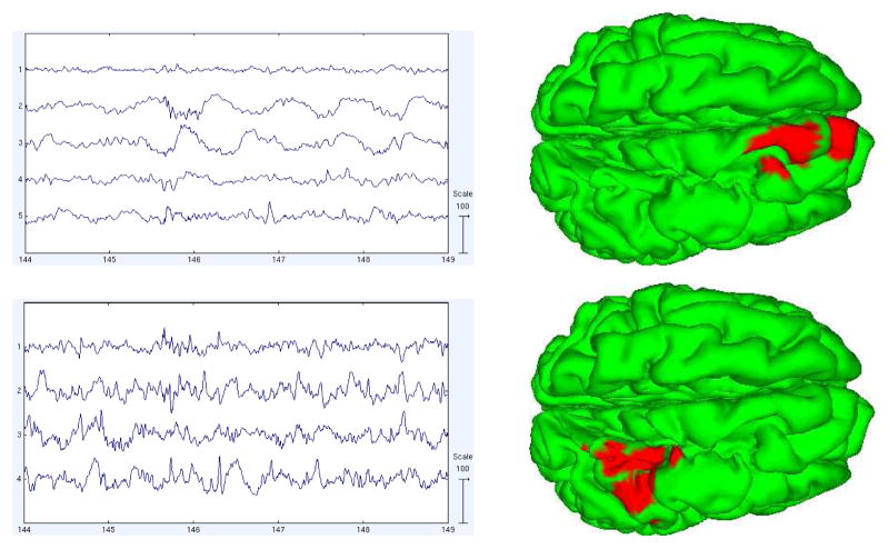
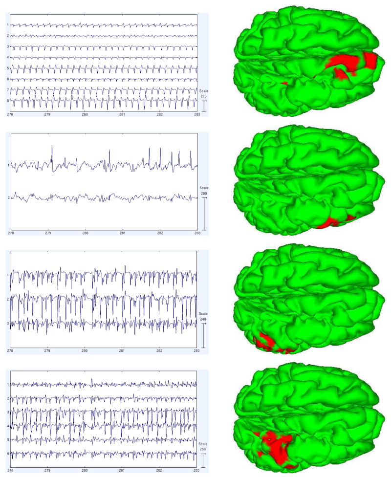
Here we report first results of numerical methods for modeling the dynamic structure and evolution of epileptic seizure activity in an intracranial subdural electrode recording from a patient with partial refractory epilepsy. A 16-min dataset containing two seizures was decomposed using up to five competing adaptive mixture independent component analysis (AMICA) models. Multiple models modeled early or late ictal, or pre- or post-ictal periods in the data, respectively. To localize sources, a realistic Boundary Element Method (BEM) head model was constructed for the patient with custom open skull and plastic (non-conductive) electrode holder features. Source localization was performed using Sparse Bayesian Learning (SBL) on a dictionary of overlapping multi-scale cortical patches constructed from 80,130 dipoles in gray matter perpendicular to the cortical surface. Remaining mutual information among seizure-model AMICA components was dominated by two dependent component subspaces with largely contiguous source domains localized to superior frontal gyrus and precentral gyrus; these accounted for most of the ictal activity. Similar though much weaker dependent subspaces were also revealed in pre-ictal data by the associated AMICA model. Electrocortical source imaging appears promising both for clinical epilepsy research and for basic cognitive neuroscience research using volunteer patients who must undergo invasive monitoring for medical purposes.
Figures
References
-
- Akalin Acar Z, Makeig S, Worrell G. Head modeling and cortical localization in epilepsy. Proc. of IEEE EMBC; 2008; Vancouver, Canada. - PubMed
-
- Makeig S, Bell AJ, Jung T-P, Sejnowski TJ. Independent component analysis of electroencephalographic data. In: Touretzky D, Mozer M, Hasselmo M, editors. Advances in Neural Information Processing Systems. Vol. 8. MIT Press; Cambridge, MA: 1996. pp. 145–151.
-
- Palmer JA, Kreutz-Delgado K, Rao BD, Makeig S. Modeling and Estimation of Dependent Subspaces. Proceedings of the 7th International Conference on Independent Component Analysis and Signal Separation; 2007.
Publication types
MeSH terms
Grants and funding
LinkOut - more resources
Full Text Sources