. 2012 Jan 18:10:3.
doi: 10.1186/1477-7827-10-3.

Prostaglandin receptors EP and FP are regulated by estradiol and progesterone in the uterus of ovariectomized rats

Affiliations

Prostaglandin receptors EP and FP are regulated by estradiol and progesterone in the uterus of ovariectomized rats

Chellakkan S Blesson et al. Reprod Biol Endocrinol. .

Abstract

Background: Prostaglandins are important for female reproduction. Prostaglandin-E2 acts via four different receptor subtypes, EP1, EP2, EP3 and EP4 whereas prostaglandin-F2alpha acts through FP. The functions of prostaglandins depend on the expression of their receptors in different uterine cell types. Our aim was to investigate the expression of EPs and FP in rat uterus and to identify the regulation by estradiol, progesterone and estrogen receptor (ER) selective agonists.

Methods: We performed four different rat experiments involving treatments with estradiol, progesterone and ER agonists. Real-time PCR and immunohistochemistry were employed to evaluate receptor expression.

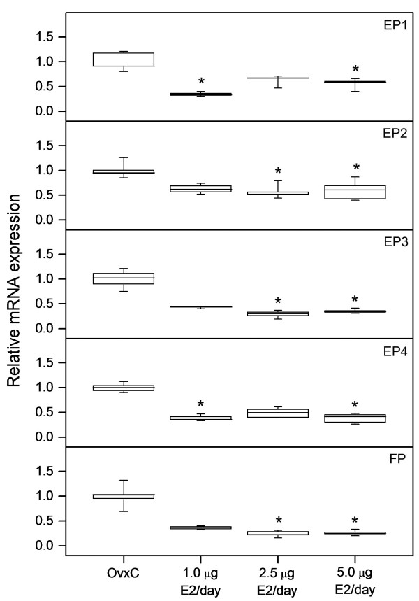
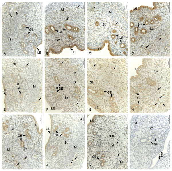
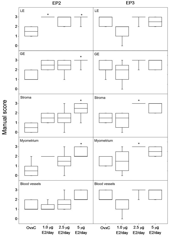
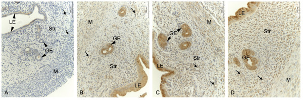
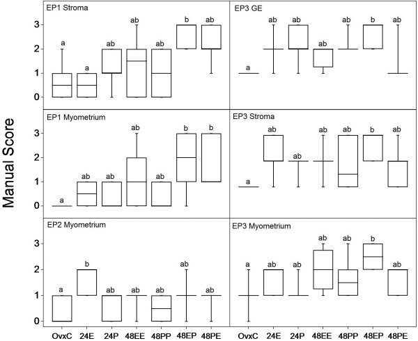
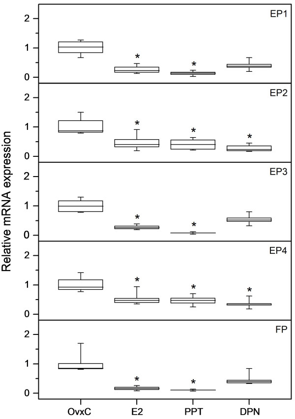
Results: Our results showed that all mRNAs and proteins of EPs and FP are expressed in the rat uterus. The expression pattern and intensity of immunostaining vary between different cell types and treatments. The mRNA expression of all EPs and FP are downregulated by estradiol and the ERalpha specific agonist PPT, whereas the ERbeta specific agonist DPN downregulates only EP2 and EP4. The protein expression however, showed an increase in EP2 and EP3 after estradiol treatment. When treated with estradiol and progesterone in combination, the expressions of EP1 and EP3 are upregulated.

Conclusions: Regulation of EPs and FP expression by estradiol appears to be mainly modulated via ERalpha for EP1, EP3 and FP, while EP2 and EP4 also are affected by the ERbeta selective ligand. Our immunohistochemical data shows a cell specific regulation of prostaglandin receptors under the influence of ovarian steroids, where EP2 is estrogen regulated in all uterine tissues examined. EP1 and EP3 are upregulated by the combination of estradiol and progesterone. Thus, our observations indicate that estradiol and progesterone regulate the mRNA and protein expression of EPs and FP in a receptor and tissue specific way.


Figures

Figure 1
Figure 1
Regulation of EP and FP mRNA by estradiol. The relative mRNA expression levels of EP1, EP2, EP3, EP4 and FP as measured by real-time PCR in uteri from ovariectomized rats treated with different doses of estradiol (E2) along with vehicle treated controls (OvxC). Box and whisker plots representing the median value with 50% of all data falling within the box. The whiskers extend to the 5th and 95th percentiles. Bars with asterisks are significantly (P < 0.05) different from OvxC.
Figure 2
Figure 2
Localization of EP and FP protein and their regulation by estradiol. Immunohistochemical localization of prostaglandin receptors in luminal epithelium (LE), glandular epithelium (GE), stroma (Str), myometrium (M) and blood vessels (arrows) in the uteri of ovariectomized controls and different doses of estradiol (E2) treated rats. Representative images of EP2 (A-D); OvxC (A), treatment with 1 μg E2 (B), 2.5 μg E2 (C) and 5 μg E2 (D), EP3 (E-H); OvxC (E), treatment with 1 μg E2 (F), 2.5 μg E2 (G) and 5 μg E2 (H). Representative images of EP1 (I), EP4 (J), FP (K) immunostaining and a negative control (L). Magnification - 200X, Scale bar = 100 μm.
Figure 3
Figure 3
Regulation of EP2 and EP3 protein by different doses of estradiol. Results from manual scoring of EP2 and EP3 immunostaining in different uterine cell types when treated with different doses of estradiol (E2). Box and whisker plots representing the median value with 50% of all data falling within the box. The whiskers extend to the 5th and 95th percentiles. Bars with asterisks are significantly (P < 0.05) different from the control group (OvxC). LE- luminal epithelium, GE- glandular epithelium.
Figure 4
Figure 4
Temporal regulation of EP2 protein by estradiol. Immunohistochemical localization of prostaglandin receptors in luminal epithelium (LE), glandular epithelium (GE), stroma (Str), myometrium (M) and blood vessels (arrows) in the uterus of ovariectomized rats. Representative images of EP2 (A-D); OvxC (A), treatment with estradiol (E2) for 1 day (B), 4 days (C) and 7 days (D). Magnification - 200X, Scale bar = 100 μm.
Figure 5
Figure 5
Cell type specific regulation of EP1, EP2 and EP3 by estradiol and progesterone. Results from manual scoring of EP1 (stroma and myometrium), EP2 (myometrium) and EP3 (glandular epithelium (GE), stroma and myometrium) when treated with estradiol (E), progesterone (P), or combinations for 24 (E or P) and 48 hours (EE, PP, EP or PE). Box and whisker plots representing the median value with 50% of all data falling within the box. The whiskers extend to the 5th and 95th percentiles. Values with different letter designations are significantly different (P < 0.05).
Figure 6
Figure 6
Downregulation of EP and FP mRNA by estradiol, PPT and DPN. The relative mRNA expression levels of EP1, EP2, EP3, EP4 and FP as measured by real-time PCR in uteri from ovariectomized rats treated with estradiol (E2), PPT or DPN along with vehicle treated controls (OvxC). Box and whisker plots representing the median value with 50% of all data falling within the box. The whiskers extend to the 5th and 95th percentiles. Bars with asterisks are significantly (P < 0.05) different from OvxC.

Similar articles

Cited by

References

    1. Catalano RD, Wilson MR, Boddy SC, Jabbour HN. Comprehensive expression analysis of prostanoid enzymes and receptors in the human endometrium across the menstrual cycle. Mol Hum Reprod. 2011;17:182–192. doi: 10.1093/molehr/gaq094. - DOI - PMC - PubMed
    1. Yang ZM, Das SK, Wang J, Sugimoto Y, Ichikawa A, Dey SK. Potential sites of prostaglandin actions in the periimplantation mouse uterus: differential expression and regulation of prostaglandin receptor genes. Biol Reprod. 1997;56:368–379. doi: 10.1095/biolreprod56.2.368. - DOI - PubMed
    1. Lim H, Dey SK. Prostaglandin E2 receptor subtype EP2 gene expression in the mouse uterus coincides with differentiation of the luminal epithelium for implantation. Endocrinology. 1997;138:4599–4606. doi: 10.1210/en.138.11.4599. - DOI - PubMed
    1. Kennedy TG. Evidence for a role for prosaglandins in the initiation of blastocyst implantation in the rat. Biol Reprod. 1977;16:286–291. doi: 10.1095/biolreprod16.3.286. - DOI - PubMed
    1. Myatt L, Lye SJ. Expression, localization and function of prostaglandin receptors in myometrium. Prostaglandins Leukot Essent Fatty Acids. 2004;70(2):137–148. doi: 10.1016/j.plefa.2003.04.004. - DOI - PubMed

Publication types

MeSH terms

LinkOut - more resources