. 2012 Feb;71(2):156-61.
doi: 10.1038/pr.2011.22. Epub 2011 Dec 21.

Changes in the frequency and in vivo vessel-forming ability of rhesus monkey circulating endothelial colony-forming cells across the lifespan (birth to aged)

Affiliations

Changes in the frequency and in vivo vessel-forming ability of rhesus monkey circulating endothelial colony-forming cells across the lifespan (birth to aged)

W Chris Shelley et al. Pediatr Res. 2012 Feb.

Abstract

Introduction: We have identified a novel hierarchy of human endothelial colony-forming cells (ECFCs) that are functionally defined by their proliferative and clonogenic potential and in vivo vessel-forming ability. The rhesus monkey provides an excellent model in which to examine the changes in circulating concentrations and functions of ECFCs since this nonhuman primate possesses a long lifespan and has been used extensively to model age-related processes that occur in humans.

Results: Endothelial cells (ECs) derived from rhesus monkey ECFCs share a cell-surface phenotype similar to human cord blood ECFCs, rapidly form capillary-like structures in vitro, and form endothelial-lined vessels in vivo upon implantation in immunodeficient mice in an age-dependent manner. Of interest, although ECFCs from the oldest monkeys formed capillary-like structures in vitro, the cells failed to form inosculating vessels when implanted in vivo and displayed a deficiency in cytoplasmic vacuolation in vitro; a critical first step in vasculogenesis.

Discussion: Utilizing previously established clonogenic assays for defining different subpopulations of human ECFCs, we have shown that a hierarchy of ECFCs, identical to human cells, can be isolated from the peripheral blood of rhesus monkeys, and that the frequency of the circulating cells varies with age. These studies establish the rhesus monkey as an important preclinical model for evaluating the role and function of circulating ECFCs in vascular homeostasis and aging.

Methods: Peripheral blood samples were collected from 40 healthy rhesus monkeys from birth to 24 years of age for ECFC analysis including immunophenotyping, clonogenic assays, and in vivo vessel formation.


Figures

Figure 1
Figure 1. Phenotypic and functional analysis of rhesus monkey peripheral blood ECFC-derived ECs
(A) Representative photomicrographs are shown of individual rhesus monkey ECFC derived EC colony from peripheral blood followed by EC colonies expanding to form an EC monolayer. (B) Immunophenotyping of cell monolayers derived from monkey peripheral blood ECFC-derived ECs by fluorescence cytometry is presented. These ECs express CD31, CD34, CD105, CD144, CD146, VEGFR2, and UEA I but not CD45, CD14, or AC133. The final panel depicts peripheral blood mononuclear (Blood Mnl) CD45 expression as a comparative control. (C) Incorporation of 488-Ac-LDL by monkey ECFC-derived ECs. These ECs were able to ingest 488-Ac-LDL (green); nuclei were stained with DAPI (blue). (D) Formation of capillary-like structures when monkey ECFC-derived ECs were plated on Matrigel. Scale bar represents 100 μm. Shown is representative data from 28 independent experiments (at least one analysis of each of 28 animals).
Figure 2
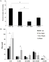
Figure 2. Quantitation of the clonogenic and proliferative potential of single ECs derived from rhesus monkey peripheral blood
(A) The percentage of single monkey ECFC-derived ECs dividing at least once after 14 days in culture is depicted. There were noticeable fewer cells undergoing division in 4-<18 yr-old animals compared to the younger animals. (B) The distribution of different size colonies derived from single ECs in an individual well after 14 days of culture is represented. There was a dramatically higher percent of cells that formed HPP-ECFC colonies in the animals <4 years of age compared to those >4 years of age. *P<0.05, by parametric ANOVA (N=8/group).
Figure 3
Figure 3. Rhesus monkey derived ECFC demonstrate the potential to form functional capillaries in vivo.
(A) Anti-CD31 labeled monkey vessels (*) containing mouse RBCs and mouse vessels (arrow head) are shown. (B) Higher magnification of anti-CD31 labeled monkey capillary containing mouse RBC. (C) Higher magnification of mouse capillary that fails to stain with the anti-CD31 antibody. Scale bar for A is 100 μm and 50 μm for B and C (representative data from 4 subjects/group implanted into a total of 16 recipient mice).
Figure 4
Figure 4. Rhesus ECFCs derived from different ages display a different ability to form vacuoles in vitro
(A) 1–4 year old and (B) > 18 year old rhesus monkey ECFC are able to form vacuoles in vitro (arrows indicate vacuoles; scale bar 100 μm). 1–4 year old rhesus ECFCs demonstrated an increased vacuole density (C), vacuole area (D), and total vacuole area (E) compared to > 18 year old Rhesus ECFCs (* denotes p-value < 0.05)(3 individuals from each group analyzed).

References

    1. Hirschi KK, Ingram DA, Yoder MC. Assessing identity, phenotype, and fate of endothelial progenitor cells. Arterioscler Thromb Vasc Biol. 2008;28:1584–1595. - PMC - PubMed
    1. Asahara T, Murohara T, Sullivan A, Silver M, van der Zee R, Li T, Witzenbichler B, Schatteman G, Isner JM. Isolation of putative progenitor endothelial cells for angiogenesis. Science. 1997;275:964–967. - PubMed
    1. Povsic TJ, Zavodni KL, Vainorius E, Kherani JF, Goldschmidt-Clermont PJ, Peterson ED. Common endothelial progenitor cell assays identify discrete endothelial progenitor cell populations. Am Heart J. 2009;157:335–344. - PMC - PubMed
    1. Timmermans F, Plum J, Yoder MC, Ingram DA, Vandekerckhove B, Case J. Endothelial progenitor cells: identity defined? J Cell Mol Med. 2009;13:87–102. - PMC - PubMed
    1. Kawamoto A, Losordo DW. Endothelial progenitor cells for cardiovascular regeneration. Trends Cardiovasc Med. 2008;18:33–37. - PMC - PubMed

Publication types

MeSH terms