. 2012 Apr;86(7):3924-33.
doi: 10.1128/JVI.06478-11. Epub 2012 Jan 18.

HIV cell-to-cell transmission requires the production of infectious virus particles and does not proceed through env-mediated fusion pores

Affiliations

HIV cell-to-cell transmission requires the production of infectious virus particles and does not proceed through env-mediated fusion pores

Blandine Monel et al. J Virol. 2012 Apr.

Abstract

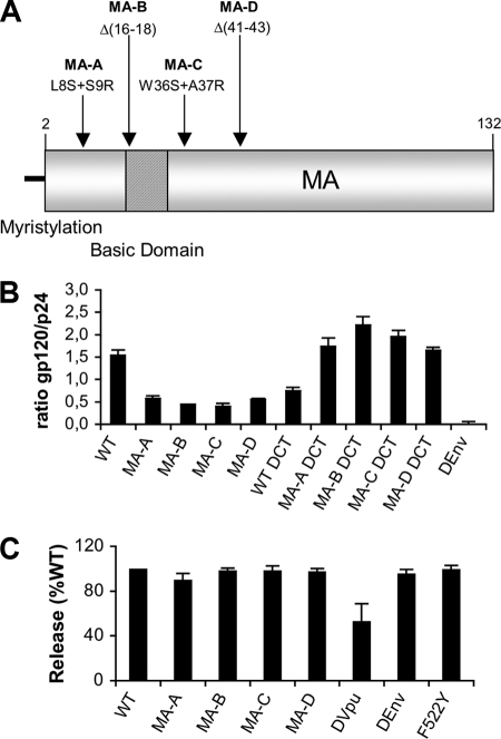
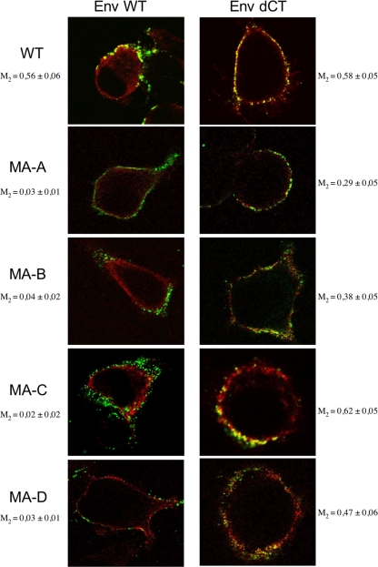
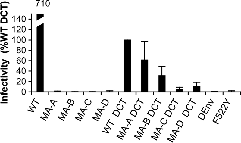
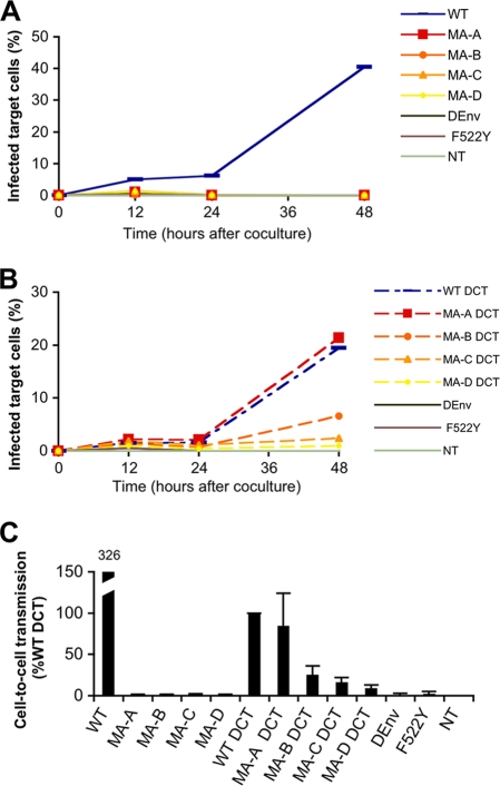
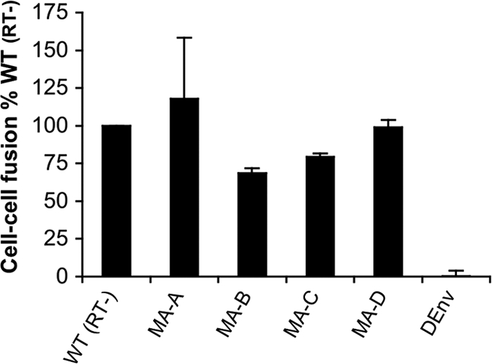
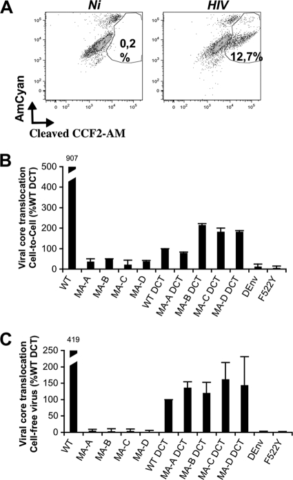
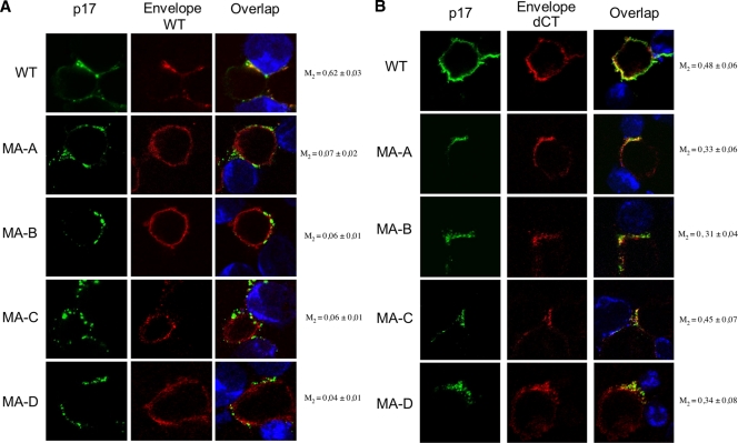
Direct cell-to-cell transmission of human immunodeficiency virus (HIV) is a more potent and efficient means of virus propagation than infection by cell-free virus particles. The aim of this study was to determine whether cell-to-cell transmission requires the assembly of enveloped virus particles or whether nucleic acids with replication potential could translocate directly from donor to target cells through envelope glycoprotein (Env)-induced fusion pores. To this end, we characterized the transmission properties of viruses carrying mutations in the matrix protein (MA) that affect the incorporation of Env into virus particles but do not interfere with Env-mediated cell-cell fusion. By use of cell-free virus, the infectivity of MA mutant viruses was below the detection threshold both in single-cycle and in multiple-cycle assays. Truncation of the cytoplasmic tail (CT) of Env restored the incorporation of Env into MA mutant viruses and rescued their cell-free infectivity to different extents. In cell-to-cell transmission assays, MA mutations prevented HIV transmission from donor to target cells, despite efficient Env-dependent membrane fusion. HIV transmission was blocked at the level of virus core translocation into the cytosol of target cells. As in cell-free assays, rescue of Env incorporation by truncation of the Env CT restored the virus core translocation and cell-to-cell infectivity of MA mutant viruses. These data show that HIV cell-to-cell transmission requires the assembly of enveloped virus particles. The increased efficiency of this infection route may thus be attributed to the high local concentrations of virus particles at sites of cellular contacts rather than to a qualitatively different transmission process.


Figures

Fig 1
Fig 1
Characterization of HIV WT and MA mutant particles. (A) Schematic representation of the HIV MA protein, showing the positions of mutated residues. (B) Incorporation of HIV Env protein into WT and MA mutant HIV-1 particles. Virus stocks of the indicated molecular clones were obtained by transfecting HeLa cells as described in Materials and Methods. The incorporation of Env into virions was assessed by using viruses purified by centrifugation through a 20% sucrose cushion. Viral pellets were lysed in TNE buffer containing 1% Triton X-100. The concentrations of p24 and of gp120 were quantified by dedicated ELISAs, and their ratios were calculated. The results shown represent the means for two independent experiments, each conducted in duplicate wells. Errors bars indicate standard deviations. The DEnv clone does not encode an Env protein. (C) Release of WT and MA mutant HIV particles into the supernatant. HeLa cells were transfected with the indicated molecular clones, and the quantities of the HIV-1 p24 protein in the supernatant and inside the cells were determined by ELISA. The results shown represent the calculated ratio of p24 in the supernatant to total p24 as a percentage of the ratio for WT HIV. The data are expressed as means ± standard deviations for four independent experiments. DVpu is a proviral clone defective for Vpu, and the F522Y mutant encodes a fusion-defective Env protein.
Fig 2
Fig 2
Analysis of the subcellular localizations of the HIV matrix protein and Env by confocal microscopy. HeLa cells transfected with the indicated molecular clones were fixed and processed for immunofluorescence analysis with anti-Env MAb 2G12 (red) and a mouse MAb specific for the mature MA protein (green). Representative confocal images for each clone are shown. The Manders overlap coefficient (M) was calculated by setting the area of interest in the green channel (MA) and analyzing 5 cells per sample (10 Z-stacks/cell).
Fig 3
Fig 3
Single-round infectivity of WT or MA mutant HIV particles in a cell-free virus assay. P4C5 cells were exposed to a virus produced in HeLa cells. Four serial dilutions of each virus stock were tested in 96-well plates. After 48 h, infection of the target cells was assessed by staining with chlorophenolred-β-d-galactopyranoside (CPRG). The infectivity of each virus is expressed as the percentage of WT DCT infectivity. The data are expressed as means ± standard deviations for three independent experiments.
Fig 4
Fig 4
Measurement of cell-to-cell transmission by flow cytometry. (A and B) HeLa cells were transfected with the indicated molecular clones. Twenty-four hours later, HeLa cells were washed to eliminate free virions, and MT4R5 target cells (labeled with CFSE) were added for 4 h at 37°C. Target cells were then collected, and the percentages of Gag+ cells were determined by flow cytometry at 15 h, 24 h, and 48 h after coculture. The results of one representative experiment are shown for clones expressing wild-type Env (A) and CT-truncated Env (B). (C) For each clone, the area under the curve was calculated and expressed as a percentage of that for the WT DCT clone. The data are expressed as means ± standard deviations for three independent experiments.
Fig 5
Fig 5
Fusion between transfected HeLa cells and JLTRG-R5 reporter cells. HeLa cells were transfected with the indicated molecular clones. The WT(RT) clone expresses wild-type MA and Env sequences, but its replication is prevented by 2 mutations in the RT catalytic site. Twenty-four hours later, HeLa cells were washed, and JLTRG-R5 target cells were added for 4 h in the presence of nevirapine, after which they were removed and cultured for 20 h. Cell-cell fusion efficiency was assessed by measuring Tat-induced LTR-GFP expression. All viral clones for which results are shown are replication defective, and the GFP signal is due only to Tat protein from donor cells that penetrates into target cells. Results are represented as the percentage of the result for the WT(RT) clone, based on three independent experiments.
Fig 6
Fig 6
Translocation of the viral core into the cytoplasm of target cells for WT HIV and MA mutants. MT4R5 cells either were cocultivated with donor HeLa cells for 2 h or were directly exposed to Vpr-βLam cell-free virus for 2 h, harvested, and incubated at room temperature for 2 h with CCF2-AM. Viral fusion was evaluated by measuring the percentage of cells positive for the cleaved form of CCF2-AM. (A) Flow cytometric analysis for one representative experiment. Ni, noninfected control cultures. (B and C) Percentages of cells positive for cleaved CCF2-AM in the context of a cell-to-cell assay (B) or a cell-free virus assay (C) carried out in three independent experiments. The data are expressed as percentages of the value for WT DCT and are means ± standard deviations.
Fig 7
Fig 7
Localization of mature HIV MA protein and WT or truncated Env in a coculture of transduced MT4R5 cells and target MT4R5 cells. MT4R5 cells were transduced with VSV-G-pseudotyped lentiviral vectors carrying the RNAs of different MA mutant clones with WT or truncated Env genes. HIV-expressing MT4R5 donor cells were mixed in a 1:1 ratio with Far Red dye-labeled recipient cells (blue) for 1 h 30 min at 37°C. Cells were then stained for MA (green) and Env (red) and were analyzed by confocal microscopy in the context of WT Env (A) or CT-truncated Env (B). The Manders overlap coefficient (M) was calculated by setting the area of interest in the green channel (MA) and analyzing at least 5 HIV-positive cells establishing contacts with neighbor cells (10 Z-stacks/cell).

Similar articles

Cited by

References

    1. Akari H, Fukumori T, Adachi A. 2000. Cell-dependent requirement of human immunodeficiency virus type 1 gp41 cytoplasmic tail for Env incorporation into virions. J. Virol. 74:4891–4893 - PMC - PubMed
    1. Beaumont E, et al. 2009. Matrix and envelope coevolution revealed in a patient monitored since primary infection with human immunodeficiency virus type 1. J. Virol. 83:9875–9889 - PMC - PubMed
    1. Berlioz-Torrent C, et al. 1999. Interactions of the cytoplasmic domains of human and simian retroviral transmembrane proteins with components of the clathrin adaptor complexes modulate intracellular and cell surface expression of envelope glycoproteins. J. Virol. 73:1350–1361 - PMC - PubMed
    1. Bhatia AK, Kaushik R, Campbell NA, Pontow SE, Ratner L. 2009. Mutation of critical serine residues in HIV-1 matrix result in an envelope incorporation defect which can be rescued by truncation of the gp41 cytoplasmic tail. Virology 384:233–241 - PMC - PubMed
    1. Bieniasz PD. 2009. The cell biology of HIV-1 virion genesis. Cell Host Microbe 5:550–558 - PMC - PubMed

Publication types

MeSH terms

Substances