. 2012 Apr;86(7):3828-38.
doi: 10.1128/JVI.06765-11. Epub 2012 Jan 18.

Mutation of the f-protein cleavage site of avian paramyxovirus type 7 results in furin cleavage, fusion promotion, and increased replication in vitro but not increased replication, tissue tropism, or virulence in chickens

Affiliations

Mutation of the f-protein cleavage site of avian paramyxovirus type 7 results in furin cleavage, fusion promotion, and increased replication in vitro but not increased replication, tissue tropism, or virulence in chickens

Sa Xiao et al. J Virol. 2012 Apr.

Abstract

We constructed a reverse genetics system for avian paramyxovirus serotype 7 (APMV-7) to investigate the role of the fusion F glycoprotein in tissue tropism and virulence. The AMPV-7 F protein has a single basic residue arginine (R) at position -1 in the F cleavage site sequence and also is unusual in having alanine at position +2 (LPSSR↓FA) (underlining indicates the basic amino acids at the F protein cleavage site, and the arrow indicates the site of cleavage.). APMV-7 does not form syncytia or plaques in cell culture, but its replication in vitro does not depend on, and is not increased by, added protease. Two mutants were successfully recovered in which the cleavage site was modified to mimic sites that are found in virulent Newcastle disease virus isolates and to contain 4 or 5 basic residues as well as isoleucine in the +2 position: (RRQKR↓FI) or (RRKKR↓FI), named Fcs-4B or Fcs-5B, respectively. In cell culture, one of the mutants, Fcs-5B, formed protease-independent syncytia and grew to 10-fold-higher titers compared to the parent and Fcs-4B viruses. This indicated the importance of the single additional basic residue (K) at position -3. Syncytium formation and virus yield of the Fcs-5B virus was impaired by the furin inhibitor decanoyl-RVKR-CMK, whereas parental APMV-7 was not affected. APMV-7 is avirulent in chickens and is limited in tropism to the upper respiratory tract of 1-day-old and 2-week-old chickens, and these characteristics were unchanged for the two mutant viruses. Thus, the acquisition of furin cleavability by APMV-7 resulted in syncytium formation and increased virus yield in vitro but did not alter virus yield, tropism, or virulence in chickens.


Figures

Fig 1
Fig 1
Schematic diagram of the full-length cDNA encoding the APMV-7 antigenomic RNA. (A) The seven cDNA fragments were inserted sequentially in pBR322/dr vector, flanked on the upstream side by a T7 RNA polymerase promoter sequence and on the downstream side by the hepatitis delta virus ribozyme sequence (Rbz) followed by a T7 terminator sequence (Tm). The unique restriction enzymes used in the assembly and their nucleotide positions in the genome are shown: the SacII, SpeI, MluI, and NotI sites were created by nucleotide substitutions in nontranslated regions, while the other sites occurred naturally. (B) Diagram of the insertion of the EGFP open reading frame (ORF) into the APMV-7 cDNA construct at the SacII site present in the downstream nontranslated region of the P gene. The DNA insert bearing the EGFP ORF contained on its upstream end a gene end (GE) signal identical to that of the P gene, an intergenic sequence (IGS) identical to that of the F and HN genes, and a gene start (GS) signal identical to that of the N gene, followed by the EGFP ORF preceded by a sequence (Kozak) designed for efficient translation. (C) Expression of the EFGP protein in Vero cells from recombinant APMV-7. The cells were infected with either rAPMV-7 or rAPMV-7/EGFP at an MOI of 0.1. The EGFP expression was observed 24 h postinfection by light microscopy in the upper panels and fluorescence microscopy in the lower panels.
Fig 2
Fig 2
APMV-7 F protein cleavage site mutants. (A) Sequences of the cleavage sites of wt APMV-7 (top line) and four cleavage site mutants. Only wt APMV-7 and the Fcs-4B and -5B mutants could be recovered in infectious virus. The basic residues are underlined, and the mutated residues are shown in bold. (B) Syncytium formation. Vero cells were infected with parental biological APMV-7 and the indicated recombinant viruses at an MOI of 0.01 and incubated for 5 days in serum-free medium. The upper row of panels shows the cultures visualized by light microscopy. The lower row of panels shows the cultures following fixation with methanol and immunoperoxidase staining using a rabbit antiserum raised against the APMV-7 N protein. The arrows indicate syncytia. (C) Plaque formation. Vero cells were infected with the indicated viruses and incubated under conditions of methylcellulose overlay in serum-free media. On day 7, the plaques were fixed and immunostained as described for panel B.
Fig 3
Fig 3
Replication kinetics of parental biological APMV-7, wt rAMPV-7, and the indicated recombinant mutant viruses in embryonated chicken eggs (A) and in Vero (B) and DF-1 (C) cells. (A) Nine-day-old SPF embryonated chicken eggs were inoculated with 103 TCID50 of the indicated viruses. Allantoic fluids were harvested daily for virus titration. (B and C) Vero (B) and DF-1 (C) cells were infected in duplicate with the indicated viruses at an MOI of 1, and samples were collected from the culture supernatant at 24-h intervals until 120 h postinfection. Virus titers of the samples were determined by serial endpoint dilution, with infected wells detected by immunoperoxidase staining using anti-APMV-7 N protein rabbit serum.
Fig 4
Fig 4
Effects of a furin inhibitor on the formation of syncytia and plaques by wt rAPMV-7 and the Fcs-5B mutant. (A) Syncytium formation. Vero cells were infected with wt rAPMV-7 and the Fcs-5B mutant at an MOI of 0.01. The cultures were incubated with serum-free media containing furin inhibitor at 0, 1, 5, and 10 μM, as indicated. The infected cells were fixed with methanol at 4 dpi and visualized by immunoperoxidase staining with anti-APMV-7 N protein rabbit serum. (B) Plaque formation. Vero cells were infected with the Fcs-5B mutant at an MOI of 0.01 and incubated under conditions of methylcellulose overlay in serum-free media containing furin inhibitor at 0, 1, 5, and 10 μM, as indicated. At 7 dpi, the cells were fixed and stained as described for panel A.
Fig 5
Fig 5
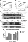
Effects of a furin inhibitor on replication of wtrAPMV-7 and the Fcs-5B mutant. Vero cells in 6-well plates were infected with wt rAPMV-7 (A and C) or the Fcs-5B mutant (B and D) at an MOI of 0.01. The cells were incubated with serum-free media containing furin inhibitor at 0, 1, 5, and 10 μM, as indicated. (A and B) Virus replication. Samples of the culture medium supernatants were collected at 24-h intervals, and virus titers were determined by limiting dilution assay. (C and D) Western blot analysis. The cells in additional plates that had been infected and treated in parallel were harvested and lysed at 2, 4, and 6 dpi. Western blot analysis was performed by using rabbit antiserum that had been raised against a synthetic peptide representing the cytoplasmic tail of the APMV-7 F protein (upper panel in C and D) or using anti-N protein rabbit serum (lower panel in C and D). The precursor (F0) and cleaved subunit (F1) of the F protein are indicated. (E) Efficiency of cleavage of the F proteins of wt rAPMV-7 and the Fcs-5B mutant in the absence of the furin inhibitor. The relative levels of the F0 and F1 proteins in the absence of inhibitor in the Western blot images in panels C and D were measured by Bio-Rad Gel Image analysis, and the efficiency of cleavage was determined by dividing the amount of F1 by the amount of F1 plus F0.
Fig 6
Fig 6
Virus titration from APMV-7-infected chickens at 2 day postinfection. Virus titers from the indicated tissues harvested on day 2 from 1-day-old and from 2-week-old chickens infected with the viruses are shown. Chickens were inoculated by the oculonasal route. Each group is represented by 3 birds for each day. Titers are shown as mean TCID50 per gram for tissues or TCID50 per milliliter for swabs.

Similar articles

Cited by

References

    1. Alexander D. 2003. Paramyxoviridae, 11th ed Iowa State University Press, Ames, IA
    1. Alexander D, Senne D. 2008. Newcastle disease and other avian paramyxovirus and pneumovirus infection, p 75–115 In Saif YM, Fadly AM, Glisson JR. (ed), Diseases of poultry. Iowa State University Press, Ames, IA
    1. Alexander DJ, Hinshaw VS, Collins MS. 1981. Characterization of viruses from doves representing a new serotype of avian paramyxoviruses. Arch. Virol. 68:265–269 - PubMed
    1. Collins MS, Bashiruddin JB, Alexander DJ. 1993. Deduced amino acid sequences at the fusion protein cleavage site of Newcastle disease viruses showing variation in antigenicity and pathogenicity. Arch. Virol. 128:363–370 - PubMed
    1. Collins PL, et al. 1995. Production of infectious human respiratory syncytial virus from cloned cDNA confirms an essential role for the transcription elongation factor from the 5′ proximal open reading frame of the M2 mRNA in gene expression and provides a capability for vaccine development. Proc. Natl. Acad. Sci. U. S. A. 92:11563–11567 - PMC - PubMed

Publication types

MeSH terms