. 2012 Apr;86(7):3795-808.
doi: 10.1128/JVI.05972-11. Epub 2012 Jan 18.

Combination of biological screening in a cellular model of viral latency and virtual screening identifies novel compounds that reactivate HIV-1

Affiliations

Combination of biological screening in a cellular model of viral latency and virtual screening identifies novel compounds that reactivate HIV-1

Edurne Gallastegui et al. J Virol. 2012 Apr.

Abstract

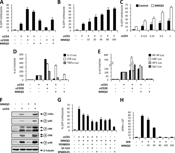
Although highly active antiretroviral therapy (HAART) has converted HIV into a chronic disease, a reservoir of HIV latently infected resting T cells prevents the eradication of the virus from patients. To achieve eradication, HAART must be combined with drugs that reactivate the dormant viruses. We examined this problem in an established model of HIV postintegration latency by screening a library of small molecules. Initially, we identified eight molecules that reactivated latent HIV. Using them as templates, additional hits were identified by means of similarity-based virtual screening. One of those hits, 8-methoxy-6-methylquinolin-4-ol (MMQO), proved to be useful to reactivate HIV-1 in different cellular models, especially in combination with other known reactivating agents, without causing T-cell activation and with lower toxicity than that of the initial hits. Interestingly, we have established that MMQO produces Jun N-terminal protein kinase (JNK) activation and enhances the T-cell receptor (TCR)/CD3 stimulation of HIV-1 reactivation from latency but inhibits CD3-induced interleukin-2 (IL-2) and tumor necrosis factor alpha (TNF-α) gene transcription. Moreover, MMQO prevents TCR-induced cell cycle progression and proliferation in primary T cells. The present study documents that the combination of biological screening in a cellular model of viral latency with virtual screening is useful for the identification of novel agents able to reactivate HIV-1. Moreover, we set the bases for a hypothetical therapy to reactivate latent HIV by combining MMQO with physiological or pharmacological TCR/CD3 stimulation.


Figures

Fig 1
Fig 1
Screening of molecules that reactivate HIV from latency in an in vitro cellular model. (A) Chemical structures of active compounds identified in a screening for HIV activation. Upon screening a library of 6,000 small molecules using a cell line containing a latent HIV-based retroviral vector (J-Lat clone A2), eight compounds were identified that reproducibly lead to an induction of HIV expression. Six of these compounds were grouped into two families according to their structural similarity, classes 1 and 2. Hits 4 and 1 are representative of these classes 1 and 2, respectively. (B) Dose response of hits 1 and 4. Cells from the J-Lat heterogeneous population were incubated with increasing concentrations of drug for 24 h and analyzed by flow cytometry. HIV-GFP reactivation is reported as the percentage of GFP-expressing cells (%GFP). Cell viability was measured by flow cytometry gating on the live population at the forward scatter (FSC) and side scatter (SSC) plot. An example is shown in panel C.
Fig 2
Fig 2
Virtual screening identifies MMQO as a novel compound that reactivates HIV from latency with no cell toxicity. (A) A library of 43 small molecules (A2 to A44) obtained by virtual screening for compounds pharmacophorically related to the initial hits was purchased and tested for HIV reactivation in a J-Lat heterogeneous cell population. Drugs were added to cells at 40 μM for 48 h, and the percentage of GFP-expressing cells (%GFP) was measured by flow cytometry. The chemical structure of the positive-hit 8-methoxy-6-methylquinolin-4-ol (MMQO) is indicated. (B) Dose response of MMQO. Cells from the J-Lat heterogeneous population were incubated with increasing concentrations of MMQO for 24 h and analyzed by flow cytometry. HIV-GFP reactivation is reported as a percentage of GFP-expressing cells (%GFP) and mean fluorescence intensity (MFI). Cell viability was measured on the FSC-SSC plot as described for Fig. 1C. Data are represented as means ± standard errors of the means (SEM) (n = 3). (C and D) Cell cycle profile of cells treated with reactivating hits. Jurkat cells were treated for 36 h with MMQO, hit 1, or hit 4 at 40 μM or were maintained untreated as a mock control, followed by propidium iodide staining and the flow cytometry analysis of the proportion of cells in each cell cycle phase. The percentage of cells in sub-G1, G1, S, and G2/M phases is shown in panel D. Data are represented as means ± SEM (n = 3). (E) Hits 1 and 4, but not MMQO, produce cell death. HeLa cells were treated for 16 h with MMQO, hit 1, or hit 4 at 80 μM or were maintained untreated as a mock control, followed by annexin V-CF647 and 7-AAD staining and the flow cytometry analysis of the proportion of cells in each staining gate. The percentage of cells in early (annexin-positive) or late (annexin-positive plus 7-AAD-positive) apoptosis is shown. Data are represented as means ± SEM (n = 3).
Fig 3
Fig 3
MMQO activates HIV at the transcriptional level independently of genome context. (A) MMQO activates latent HIV in different genomic contexts. Cells of latently infected clones J-Lat E27, A2, and H2 were incubated with increasing concentrations of MMQO for 24 h and analyzed by flow cytometry for GFP-expressing cells and cell viability. (B) MMQO activates a transfected HIV-1 LTR-luciferase reporter. 293T cells were transfected with an HIV-1 LTR-luciferase reporter plasmid. One day after transfection, cells were treated with MMQO (80 μM), EGF (50 ng/ml), PMA (10 nM), TSA (400 nM), or TNF-α (10 ng/ml) for 36 h, followed by cell lysis, protein normalization (5 μg), and luciferase activity assay. Data are expressed as relative light units (RLU). (C) Time course response of HIV-1 transcription to MMQO analyzed by RT-qPCR. J-Lat E27 cells were treated with 80 μM MMQO for the time indicated, and RNA was extracted. PMA (10 nM) and TNF-α (10 ng/ml) treatments for 12 h were added as controls. HIV transcription was measured by RT-qPCR using primers corresponding to the HIV 5′LTR (R-gag) and normalizing with GAPDH. The fold change (FC) between treated and untreated cells is shown. (D) MMQO stimulates HIV transcription initiation. J-Lat E27 cells were untreated or were treated with 80 μM MMQO for 24 h, RNA was extracted, and HIV reactivation was measured by RT-qPCR. Amplicons corresponding to the 5′LTR (R-gag), Tat, GFP, and 3′LTR (U3) were used. GAPDH expression was measured for normalization. To compare between different amplicons, qPCR was performed in parallel from genomic DNA (gDNA). Data are expressed as relative units (RU) (cDNA amplification/GAPDH)/gDNA amplification in a log scale. A scheme of the HIV minigenome with positions of qPCR primers is shown below. Throughout the figure data are represented as means ± SEM (n = 3).
Fig 4
Fig 4
Synergistic activation of HIV expression by combination of MMQO with small molecules known to reactivate latent HIV. (A) MMQO synergizes with TNF-α and PMA on the activation of a latent HIV-1 minigenome. Cells from clone J-Lat E27 were incubated for 36 h with combinations of MMQO (80 μM), TSA (400 nM), PMA (10 nM), TNF-α (10 ng/ml), and HMBA (10 mM) and analyzed by flow cytometry. HIV-GFP reactivation is reported as MFI or as the percentage of GFP-expressing cells for a selection of the treatments together with the percentage of cell viability. (B) MMQO synergizes with prostratin. J-Lat E27 cells were incubated for 36 h with combinations of MMQO (80 μM) and prostratin (2 μM) and analyzed as described for panel A. Data are represented as means ± SEM (n = 3). (C) Combined activation of clone J-Lat H2. Cells from clone J-Lat H2 were treated for 36 h with PMA (10 nM), TSA (400 nM), TNF-α (10 ng/ml), HMBA (10 mM), or 5-azadC (5 μM) in the absence or presence of MMQO (80 μM). HIV-GFP reactivation is reported as MFI. (D) Activation of HIV-1 with low doses of MMQO and PMA. J-Lat E27 cells were incubated for 16 h with combinations of suboptimal doses of MMQO and PMA and analyzed as described for panel A. Data are represented as means ± SEM (n = 3). (E) J-Lat E27 cells were treated with increasing concentrations of TNF-α (100 to 1,000 ng/ml) or PMA (10 to 160 nM) in the absence or presence (50 or 100 μM) of MMQO. HIV-GFP reactivation is reported as MFI. (F) J-Lat E27 cells were treated with increasing concentrations of MMQO (50 to 150 nM) in the absence or presence of HMBA(10 or 20 mM) or TSA (0.4 or 1 μM). HIV-GFP reactivation is reported as MFI. (G) Combinatorial treatment with MMQO and hit 1 or 4. J-Lat E27 cells were treated with increasing concentrations of hit 1 or 4 (5 to 80 μM) in the absence or presence (50 or 100 μM) of MMQO. HIV-GFP reactivation is reported as MFI. Cell viability extracted from the FSC-SSC plots is shown in the lower panels. (H) Combinatorial treatment with hits 1 and 4 and MMQO. J-Lat E27 cells were treated with increasing concentrations of hit 1 or 4 (10 to 40 μM) in the absence or presence (80 μM) of MMQO. HIV-GFP reactivation is reported as MFI.
Fig 5
Fig 5
MMQO reactivates latent HIV from different HIV latency models containing a full-length provirus. (A) MMQO synergizes with treatments that activate full-length HIV-1 J-Lat cells. Cells from clone J-Lat 15.4 were incubated for 36 h with combinations of TNF-α (10 ng/ml), TSA (400 nM), and 5-azadC (5 μM) in the absence or presence of MMQO (80 μM) and analyzed by flow cytometry. 5-azadC was added 24 h before other treatments (preincubation). HIV-GFP reactivation is reported as a percentage of GFP-expressing cells. (B) HIV reactivation from latently infected cell lines U1 and ACH2. U1 and ACH2 cells were incubated for 6 h with MMQO (80 μM), PMA (10 nM), TSA (400 nM), TNF-α (10 ng/ml), HMBA (10 mM), or 5-azadC (5 μM) as indicated, and RNA was extracted to assess HIV expression by RT-qPCR using primers corresponding to HIV 5′LTR (R-gag) and normalizing with GAPDH. Data are represented relative to the maximal activation for each cell line. (C) MMQO enhances HIV reactivation by TNF-α and 5-azadC in cell lines U1 and ACH2. U1 and ACH2 cells were incubated for 6 h with TNF-α (10 ng/ml) and 5-azadC (5 μM) or left untreated and were in the absence or presence of MMQO (80 μM), and RNA was extracted to assess HIV expression by RT-qPCR using primers corresponding to the HIV 5′LTR (R-gag) and normalizing with GAPDH. Data are represented as fold increases compared to the expression of untreated cells. (D) MMQO reactivation of HIV-1 from patients. Twenty to 30 million PBMC were obtained from several HAART-treated patients (named A to I) and incubated with 80 μM MMQO for 36 h or left untreated. RNA was extracted and HIV expression measured by RT-qPCR using primers corresponding to the HIV 5′LTR (R-gag) and normalizing with GAPDH. Data are expressed as relative units (RU) of HIV/GAPDH expression. (E) MMQO induces viral particle formation in ACH2 cells. ACH2 cells were incubated for 24 h with MMQO (80 μM), PMA, ionomycin (IO), or prostratin. Cell culture supernatant media were collected 24 and 72 h after the initial addition of drugs and assayed for the Gag-derived p24 HIV protein using an ELISA to measure the production of viral particles. (F) Viral particles induced in MMQO-treated ACH2 cells are infectious. Human PBMC or ACH2 cells were incubated for 24 h with MMQO (80 μM) or prostratin (2 μM), supernatant media were collected, and viral infectivity was measured using TZM-bl indicator cells harboring an LTR-luciferase reporter system. After 72 h of incubation, cell-associated luciferase activity was determined. Data are expressed as the fold increase of treated versus untreated cells in relative luciferase units (RLU). Throughout the figure data are represented as means ± SEM (n = 3).
Fig 6
Fig 6
MMQO enhances TCR/CD3 stimulation of HIV-1 reactivation from latency and prevents TCR-induced proliferation in primary T cells. (A and B) MMQO synergizes with CD3 to reactivate HIV-1 from latency. Jurkat-LAT-GFP cells were stimulated for 18 h on 96-well plates with coated anti-CD3 (1 μg/ml), soluble anti-CD28 (0.5 μg/ml), and MMQO (160 μM) separately or in combination. (B) Jurkat-LAT-GFP cells were stimulated with coated anti-CD3 and increasing concentrations of MMQO as indicated for 18 h. GFP expression was measured by flow cytometry, and results indicate the percentage of GFP+ cells. (C) Jurkat-LAT-GFP cells were stimulated with increasing concentrations of coated anti-CD3 and MMQO at 80 μM for 18 h. (D) Differential effects of MMQO on IL-2 and TNF-α promoters and HIV-LTR transactivation. Jurkat cells were transiently transfected with the plasmids IL-2-Luc, TNF-α-Luc, and LTR-Luc, and 24 h later they were stimulated with coated anti-CD3 (1 μg/ml) in the absence or the presence of either soluble anti-CD28 (0.5 μg/ml) or MMQO (160 μM) or with MMQO alone for 24 h. The results are expressed as the percentage of activation considering CD3-induced transactivation as 100% activation. (E) Activity of MMQO on NF-κB-, NFAT-, AP-1-, and Sp1-responsive minimal promoters. Jurkat cells were transfected with NF-κB, NFAT, AP-1, and Sp1 luciferase reporter plasmid DNA and stimulated as described for panel C. Luciferase activity was measured 24 h later after protein normalization. The results are expressed as the percentage of activation considering CD3-induced transactivation as 100% activation. (F) Effect of MMQO on α-CD3-induced NF-κB and MAPK activation. Jurkat cells were stimulated with anti-CD3 and MMQO (160 μM) separately or in combination for 15 min, and the levels of phospho-IκBα, phosho-p65 (Ser536), phospho-JNK, phospho-ERK, and phospho-p38 were assessed by immunoblotting with specific antibodies and anti-tubulin MAb as a loading control. (G) MMQO antagonizes HIV-1 latency through the JNK pathway. Jurkat-LAT-GFP cells were treated with the ERK inhibitor PD98059 (25 μM), IKK2 inhibitor (25 μM), and JNK inhibitor SP600125 (1 μM) or left untreated, and 30 min later they were stimulated with anti-CD3 plus MMQO (80 μM) or with MMQO alone for 18 h. HIV-GFP reactivation was assessed by flow cytometry, and results indicate the percentage of GFP+ cells. (H) Effects of MMQO on T-cell proliferation. Human PBMCs were stimulated with SEB (1 μg/ml) in the presence or the absence of increasing concentrations of MMQO for 72 h. [3H]thymidine incorporation was measured by liquid scintillation counting and is represented as disintegrations per minute (DPM). Throughout the figure data are represented as means ± SEM (n = 3).

Similar articles

Cited by

References

    1. Archin NM, et al. 2009. Expression of latent HIV induced by the potent HDAC inhibitor suberoylanilide hydroxamic acid. AIDS Res. Hum. Retrovir. 25:207–212 - PMC - PubMed
    1. Areias FM, et al. 2010. In silico directed chemical probing of the adenosine receptor family. Bioorg. Med. Chem. 18:3043–3052 - PubMed
    1. Bisgrove D, Lewinski M, Bushman F, Verdin E. 2005. Molecular mechanisms of HIV-1 proviral latency. Expert Rev. Anti Infect. Ther. 3:805–814 - PubMed
    1. Bisgrove DA, Mahmoudi T, Henklein P, Verdin E. 2007. Conserved P-TEFb-interacting domain of BRD4 inhibits HIV transcription. Proc. Natl. Acad. Sci. U. S. A. 104:13690–13695 - PMC - PubMed
    1. Blazkova J, et al. 2009. CpG methylation controls reactivation of HIV from latency. PLoS Pathog. 5:e1000554. - PMC - PubMed

Publication types

MeSH terms

Substances