

Nonstructural protein 2 of porcine reproductive and respiratory syndrome virus inhibits the antiviral function of interferon-stimulated gene 15
- PMID: 22258253
- PMCID: PMC3302520
- DOI: 10.1128/JVI.06466-11
Nonstructural protein 2 of porcine reproductive and respiratory syndrome virus inhibits the antiviral function of interferon-stimulated gene 15
Abstract
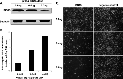
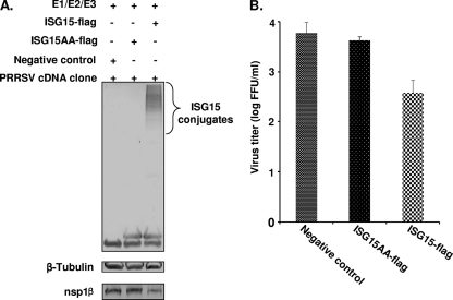
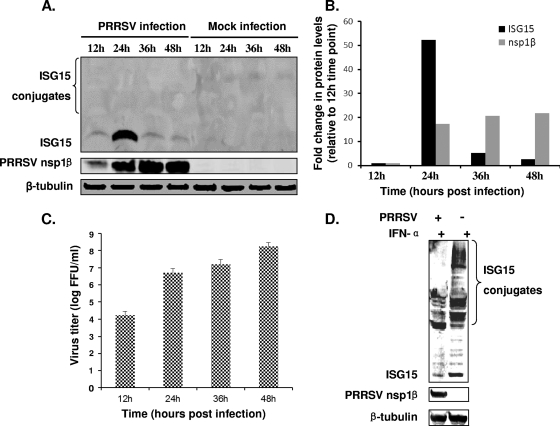
Type I interferon (alpha/beta interferon [IFN-α/β]) stimulates the expression of interferon-stimulated gene 15 (ISG15), which encodes a ubiquitin-like protein, ISG15. Free ISG15 and ISG15 conjugates function in diverse cellular pathways, particularly regulation of antiviral innate immune responses. In this study, we demonstrate that ISG15 overexpression inhibits porcine reproductive and respiratory syndrome virus (PRRSV) replication in cell culture and that the antiviral activity of interferon is reduced by inhibition of ISG15 conjugation. PRRSV nonstructural protein 2 (nsp2) was previously identified as a potential antagonist of ISG15 production and conjugation. The protein contains a papain-like protease domain (PLP2) that plays a crucial role in the proteolytic cleavage of the PRRSV replicase polyproteins. PLP2 was also proposed to belong to the ovarian tumor domain-containing superfamily of deubiquitinating enzymes (DUBs), which is capable of inhibiting ISG15 production and counteracting ISG15 conjugation to cellular proteins. To determine whether this immune antagonist function could be selectively inactivated, we engineered a panel of mutants with deletions and/or mutations at the N-terminal border of the nsp2 PLP2-DUB domain. A 23-amino-acid deletion (amino acids 402 to 424 of the ORF1a-encoded protein) largely abolished the inhibitory effect of nsp2 on ISG15 production and conjugation, but no viable recombinant virus was recovered. A 19-amino-acid deletion (amino acids 402 to 420), in combination with a downstream point mutation (S465A), partially relieved the ISG15 antagonist function and yielded a viable recombinant virus. Taken together, our data demonstrate that ISG15 and ISGylation play an important role in the response to PRRSV infection and that nsp2 is a key factor in counteracting the antiviral function of ISG15.
Figures
Similar articles
-
PRRSV nonstructural protein 11 degrades swine ISG15 by its endoribonuclease activity to antagonize antiviral immune response.Vet Microbiol. 2023 May;280:109720. doi: 10.1016/j.vetmic.2023.109720. Epub 2023 Mar 11. Vet Microbiol. 2023. PMID: 36921497
-
The Papain-Like Protease of Porcine Reproductive and Respiratory Syndrome Virus Impedes STING Translocation from the Endoplasmic Reticulum to the Golgi Apparatus by Deubiquitinating STIM1.J Virol. 2023 Apr 27;97(4):e0018823. doi: 10.1128/jvi.00188-23. Epub 2023 Apr 11. J Virol. 2023. PMID: 37039642 Free PMC article.
-
Porcine reproductive and respiratory syndrome virus nonstructural protein 4 antagonizes beta interferon expression by targeting the NF-κB essential modulator.J Virol. 2014 Sep;88(18):10934-45. doi: 10.1128/JVI.01396-14. Epub 2014 Jul 9. J Virol. 2014. PMID: 25008936 Free PMC article.
-
In vivo growth of porcine reproductive and respiratory syndrome virus engineered nsp2 deletion mutants.Virus Res. 2010 Dec;154(1-2):77-85. doi: 10.1016/j.virusres.2010.07.024. Epub 2010 Jul 29. Virus Res. 2010. PMID: 20673840 Free PMC article. Review.
-
Non-structural protein 2 of the porcine reproductive and respiratory syndrome (PRRS) virus: a crucial protein in viral pathogenesis, immunity and diagnosis.Res Vet Sci. 2013 Aug;95(1):1-7. doi: 10.1016/j.rvsc.2013.03.015. Epub 2013 Apr 13. Res Vet Sci. 2013. PMID: 23591056 Review.
Cited by
-
3Cpro of foot-and-mouth disease virus antagonizes the interferon signaling pathway by blocking STAT1/STAT2 nuclear translocation.J Virol. 2014 May;88(9):4908-20. doi: 10.1128/JVI.03668-13. Epub 2014 Feb 19. J Virol. 2014. PMID: 24554650 Free PMC article.
-
Viral OTU deubiquitinases: a structural and functional comparison.PLoS Pathog. 2014 Mar 27;10(3):e1003894. doi: 10.1371/journal.ppat.1003894. eCollection 2014 Mar. PLoS Pathog. 2014. PMID: 24676359 Free PMC article. Review.
-
The nsp2 Hypervariable Region of Porcine Reproductive and Respiratory Syndrome Virus Strain JXwn06 Is Associated with Viral Cellular Tropism to Primary Porcine Alveolar Macrophages.J Virol. 2019 Nov 26;93(24):e01436-19. doi: 10.1128/JVI.01436-19. Print 2019 Dec 15. J Virol. 2019. PMID: 31554681 Free PMC article.
-
Hyper-phosphorylation of nsp2-related proteins of porcine reproductive and respiratory syndrome virus.Virology. 2020 Apr;543:63-75. doi: 10.1016/j.virol.2020.01.018. Epub 2020 Feb 4. Virology. 2020. PMID: 32174300 Free PMC article.
-
Equine arteritis virus does not induce interferon production in equine endothelial cells: identification of nonstructural protein 1 as a main interferon antagonist.Biomed Res Int. 2014;2014:420658. doi: 10.1155/2014/420658. Epub 2014 May 25. Biomed Res Int. 2014. PMID: 24967365 Free PMC article.
References
-
- Bautista EM, Meulenberg JJ, Choi CS, Molitor TW. 1996. Structural polypeptides of the American (VR-2332) strain of porcine reproductive and respiratory syndrome virus. Arch. Virol. 141:1357–1365 - PubMed
-
- Blomstrom DC, Fahey D, Kutny R, Korant BD, Knight E., Jr 1986. Molecular characterization of the interferon-induced 15-kDa protein. Molecular cloning and nucleotide and amino acid sequence. J. Biol. Chem. 261:8811–8816 - PubMed
Publication types
MeSH terms
Substances
LinkOut - more resources
Full Text Sources
Other Literature Sources
Molecular Biology Databases
Miscellaneous
