. 2012 Apr;86(7):3647-57.
doi: 10.1128/JVI.06549-11. Epub 2012 Jan 18.

West nile virus infections suppress early viral RNA synthesis and avoid inducing the cell stress granule response

Affiliations

West nile virus infections suppress early viral RNA synthesis and avoid inducing the cell stress granule response

S C Courtney et al. J Virol. 2012 Apr.

Abstract

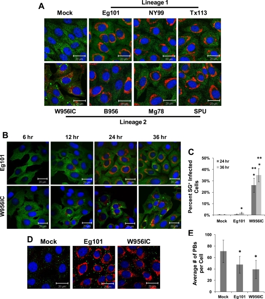
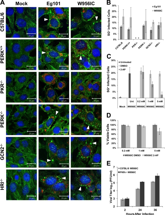
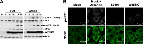
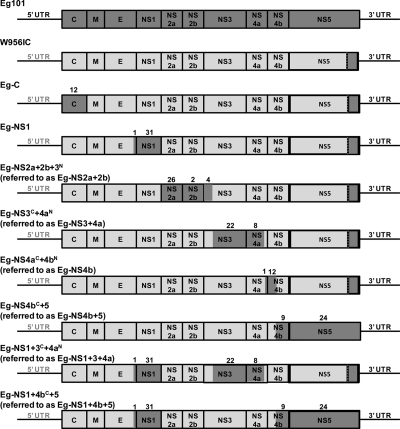
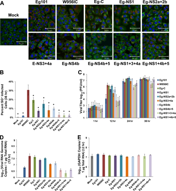
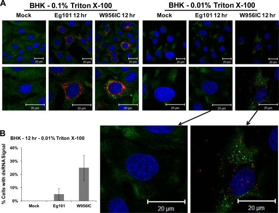
West Nile virus (WNV) recently became endemic in the United States and is a significant cause of human morbidity and mortality. Natural WNV strain infections do not induce stress granules (SGs), while W956IC (a lineage 2/1 chimeric WNV infectious clone) virus infections produce high levels of early viral RNA and efficiently induce SGs through protein kinase R (PKR) activation. Additional WNV chimeric viruses made by replacing one or more W956IC genes with the lineage 1 Eg101 equivalent in the W956IC backbone were analyzed. The Eg-NS4b+5, Eg-NS1+3+4a, and Eg-NS1+4b+5 chimeras produced low levels of viral RNA at early times of infection and inefficiently induced SGs, suggesting the possibility that interactions between viral nonstructural proteins and/or between viral nonstructural proteins and cell proteins are involved in suppressing early viral RNA synthesis and membrane remodeling during natural WNV strain infections. Detection of exposed viral double-stranded RNA (dsRNA) in W956IC-infected cells suggested that the enhanced early viral RNA synthesis surpassed the available virus-induced membrane protection and allowed viral dsRNA to activate PKR.


Figures

Fig 1
Fig 1
Stress granule induction by different strains of WNV. (A) BHK cells were infected with a lineage 1 or lineage 2 WNV strain at an MOI of 1, fixed at 36 h after infection, and analyzed by confocal microscopy. Anti-G3BP antibody (green) was used to detect SGs, anti-dsRNA antibody (red) to detect WNV-infected cells, and Hoechst dye (blue) to detect nuclei. Merged images are shown. (B) BHK cells were infected with either Eg101 (lineage 1) or W956IC (lineage 2/1-based infectious clone) virus at an MOI of 1 and analyzed for SGs and dsRNA at the indicated times after infection as described for panel A. Merged images are shown. (C) The total number of infected cells and the number of SG-positive, infected cells were counted, and the percentage of Eg101- or W956IC-infected cells containing SGs was determined as described in Materials and Methods. *, significance compared to mock-infected cells at a 95% confidence interval (P < 0.05); **, significance compared to Eg101-infected cells at a 95% confidence interval (P < 0.05). (D) BHK cells were infected with either Eg101 or W956IC at an MOI of 1 and analyzed for PBs. Anti-Dcp1a antibody (green) was used to detect PBs, anti-dsRNA antibody (red) to detect virus-infected cells, and Hoechst dye (blue) to detect nuclei. Merged images are shown. (E) PBs were counted to determine the average number per infected cell. *, significance compared to mock-infected cells at a 95% confidence interval (P < 0.05). Bars indicate ± standard deviations (SD).
Fig 2
Fig 2
Stress granule formation induced by WNV infection is PKR dependent. (A) Control MEFs (C57BL/6 and PERK+/+) and eIF2α kinase knockout MEFs (PKR−/−, PERK−/−, GCN2−/−, and HRI−/−) were infected with WNV Eg101 or W956IC at an MOI of 1, fixed at 24 h after infection, and analyzed by confocal microscopy. SGs were detected with anti-G3BP antibody (green), WNV-infected cells with anti-dsRNA antibody (red), and nuclei with Hoechst dye (blue). Arrowheads indicate SG-positive cells. Merged images are shown. (B) The total number of infected cells and the number of SG-positive, infected cells were counted, and the percentage of infected cells containing SGs was determined as described in Materials and Methods. *, significance compared to PERK+/+ control cells for the indicated infection at a 95% confidence interval (P < 0.05). (C) BHK cells were treated with different concentrations of 2-AP or DMSO (vehicle control) starting immediately after a 1-h adsorption of W956IC virus (MOI of 1), fixed at 24 h after infection, and analyzed by confocal microscopy for SGs and dsRNA as described for panel A. The percentage of SG-positive, infected cells was determined. (D) An MTT assay, performed as described in Materials and Methods, was used to assess the viability of BHK cells infected with W956IC and treated with different concentrations of 2-AP or DMSO. (E) Wild-type C57BL/6 and PKR−/− MEFs were infected with W956IC (MOI of 1). Virus yields were measured by plaque assay in BHK cells at the indicated times after infection. Bars indicate ± SD.
Fig 3
Fig 3
W956IC infection induces PKR and eIF2α phosphorylation. (A) Western blot analysis of BHK cells infected with WNV Eg101 or W956IC at an MOI of 1. Lysates were collected in radioimmunoprecipitation assay (RIPA) buffer at the indicated times (hours) after infection and analyzed using antibodies to phosphorylated PKR (Thr541), PKR, phosphorylated eIF2α (Ser51), eIF2α, and actin. M, mock. (B) BHK cells were infected with WNV Eg101 or W956IC at an MOI of 1, fixed at 24 h after infection, and analyzed by confocal microscopy. Anti-G3BP antibody (green) was used to detect SGs, and anti-p-eIF2α antibody (white) was used to detect p-eIF2α. More than 95% of the cells were infected.
Fig 4
Fig 4
Schematic representation of the WNV W956IC-Eg101 chimera genomes. Eg101 sequences are indicated by dark-gray and W956IC sequences by light-gray shading. The majority of the W956IC genome sequence is from 956D117B3, but the C terminus of NS5 and the 3′ UTR are from Eg101 (56). Additional chimeric viruses were created by replacing one or more W956IC gene regions with the Eg101 equivalent on the W956IC backbone. The number of amino acid differences between Eg101 and W956IC is indicated above each replaced gene segment.
Fig 5
Fig 5
Higher levels of early viral RNA synthesis correlate with increased early virus yields and more SG-positive, infected cells. (A) BHK cells were infected with a chimeric virus at an MOI of 1, fixed at 36 h after infection, and analyzed by confocal microscopy. SGs were detected with anti-G3BP antibody (green), WNV-infected cells with anti-dsRNA antibody (red), and nuclei with Hoechst dye (blue). Merged images are shown. (B) The total number of infected cells and the number of SG-positive, infected cells were counted, and the percentage of infected cells containing SGs was determined as described in Materials and Methods. *, significance compared to W956IC at a 95% confidence interval (P < 0.05). (C) BHK cells were infected with a chimeric virus at an MOI of 1, and virus yield was determined by plaque assay at the indicated times after infection. (D) BHK cells were infected with a chimeric virus at an MOI of 1, and total cell RNA was extracted and purified at 12 h after infection. The number of viral genome copies in each sample was determined by real-time RT-PCR using probes targeting the viral NS1 gene as described in Materials and Methods. (E) The amount of glyceraldehyde-3-phosphate dehydrogenase (GAPDH) RNA in each sample was measured by real-time RT-PCR as an internal control for sample variation. Bars indicate ± SD.
Fig 6
Fig 6
Exposed viral dsRNA was detected in 0.01% Triton X-100-permeabilized W956IC-infected, but not Eg101-infected, cells. (A) BHK cells were infected with WNV Eg101 or W956IC at an MOI of 5, fixed at 12 h after infection, permeabilized with either 0.1% or 0.01% Triton X-100, and analyzed by confocal microscopy. Anti-dsRNA antibody (red) was used to detect viral dsRNA, anti-G3BP antibody (green) to detect SGs, and Hoechst dye (blue) to detect nuclei. Merged images are shown. (B) The total number of cells and the number of dsRNA signal-positive cells from 0.01% Triton X-100-treated cells were counted, and the percentage of cells containing detectable dsRNA was determined. Bars indicate ± SD.

Similar articles

Cited by

References

    1. Ambrose RL, Mackenzie JM. 2011. West Nile virus differentially modulates the unfolded protein response to facilitate replication and immune evasion. J. Virol. 85:2723–2732 - PMC - PubMed
    1. Anderson P, Kedersha N. 2008. Stress granules: the Tao of RNA triage. Trends Biochem. Sci. 33:141–150 - PubMed
    1. Anderson P, Kedersha N. 2002. Stressful initiations. J. Cell Sci. 115:3227–3234 - PubMed
    1. Andzhaparidze OG, Bogomolova NN, Boriskin YS, Bektemirova MS, Drynov ID. 1981. Comparative study of rabies virus persistence in human and hamster cell lines. J. Virol. 37:1–6 - PMC - PubMed
    1. Bakonyi T, Hubalek Z, Rudolf I, Nowotny N. 2005. Novel flavivirus or new lineage of West Nile virus, central Europe. Emerg. Infect. Dis. 11:225–231 - PMC - PubMed

Publication types