. 2012 Mar;95(3):670-7.
doi: 10.3945/ajcn.111.026708. Epub 2012 Jan 18.

Monitoring of gluten-free diet compliance in celiac patients by assessment of gliadin 33-mer equivalent epitopes in feces

Affiliations

Monitoring of gluten-free diet compliance in celiac patients by assessment of gliadin 33-mer equivalent epitopes in feces

Isabel Comino et al. Am J Clin Nutr. 2012 Mar.

Abstract

Background: Certain immunotoxic peptides from gluten are resistant to gastrointestinal digestion and can interact with celiac-patient factors to trigger an immunologic response. A gluten-free diet (GFD) is the only effective treatment for celiac disease (CD), and its compliance should be monitored to avoid cumulative damage. However, practical methods to monitor diet compliance and to detect the origin of an outbreak of celiac clinical symptoms are not available.

Objective: We assessed the capacity to determine the gluten ingestion and monitor GFD compliance in celiac patients by the detection of gluten and gliadin 33-mer equivalent peptidic epitopes (33EPs) in human feces.

Design: Fecal samples were obtained from healthy subjects, celiac patients, and subjects with other intestinal pathologies with different diet conditions. Gluten and 33EPs were analyzed by using immunochromatography and competitive ELISA with a highly sensitive antigliadin 33-mer monoclonal antibody.

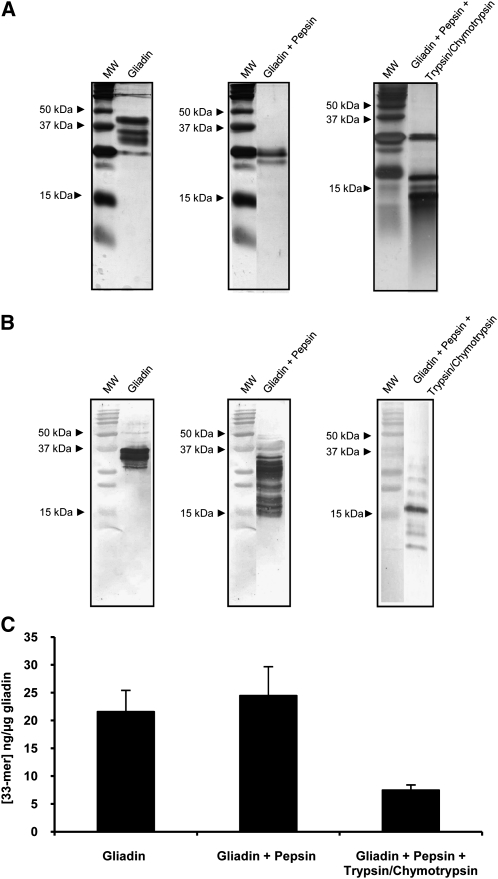
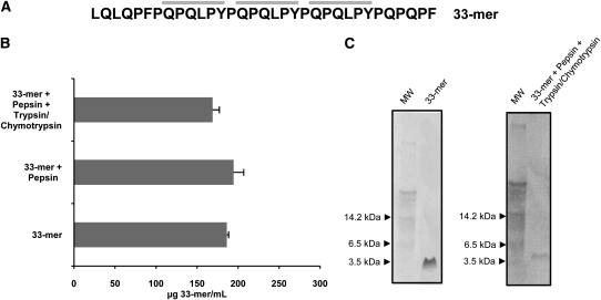
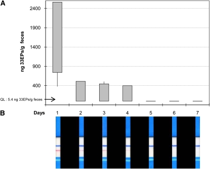
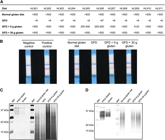
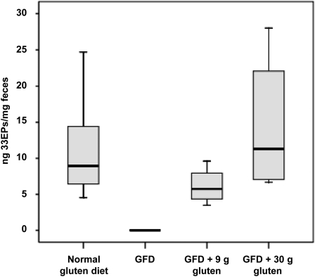
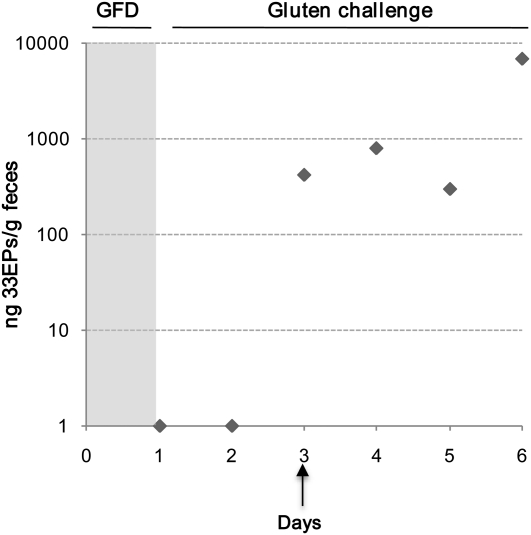
Results: The resistance of a significant part of 33EPs to gastrointestinal digestion was shown in vitro and in vivo. We were able to detect gluten peptides in feces of healthy individuals after consumption of a normal gluten-containing diet, after consumption of a GFD combined with controlled ingestion of a fixed amount of gluten, and after ingestion of <100 mg gluten/d. These methods also allowed us to detect GFD infringement in CD patients.

Conclusions: Gluten-derived peptides could be sensitively detected in human feces in positive correlation with the amount of gluten intake. These techniques may serve to show GFD compliance or infringement and be used in clinical research in strategies to eliminate gluten immunotoxic peptides during digestion. This trial was registered at clinicaltrials.gov as NCT01478867.


Figures

FIGURE 1.
FIGURE 1.
The mean (±SD) relative affinity of G12 moAb to immunotoxic peptides derived from PWG gliadin after simulated gastrointestinal digestion. A and B: SDS-PAGE and Western blot of PWG gliadin, PWG gliadin + pepsin, and PWG gliadin + pepsin + trypsin/chymotrypsin. Samples were stained with silver (A) or after transfer to a polyvinylidene fluoride membrane and incubated with G12 moAbs (B). C: Competition assay that measured the affinity of the horseradish peroxidase–conjugated G12 moAb to gliadin peptides present in gastrointestinal digests. Three assays were performed with samples run in triplicate. moAb, monoclonal antibody; MW, molecular weight marker; PWG, Prolamin Working Group.
FIGURE 2.
FIGURE 2.
Mean (±SD) resistance of 33-mer peptides to cleavage by gastrointestinal enzymes. A: Amino acid sequences of the 33-mer peptide. The G12 moAb recognition sequence in the 33-mer peptide is in bold-face type. B: Competition assay for the detection of 33-mer peptide by G12 moAb after treatment with pepsin, trypsin, and chymotrypsin. Three assays were performed with samples run in triplicate. C: Western blot analysis of 33-mer peptide by the G12 moAb after treatment with gastrointestinal enzymes. moAb, monoclonal antibody; MW, molecular weight marker.
FIGURE 3.
FIGURE 3.
Determination of the time to elimination of ingested gluten in healthy individuals subjected to a gluten-free diet for 7 d. A: Competitive assay to determine the concentration of 33EPs (ng/g) in feces (n = 4). Each sample was analyzed in triplicate, and maximum, minimum, and 25th and 75th percentiles are shown. B: One representative immunochromatographic-strip example of the trial performed with the samples collected during the study period of one subject. Blue stripes represent an internal positive control that indicates that the stick worked properly; pink stripes indicate the presence of gluten. QL, quantification limit; 33EPs, 33-mer equivalent peptidic epitopes.
FIGURE 4.
FIGURE 4.
Detection of gluten in feces of healthy individuals subjected to a controlled gluten-containing diet. A: Semiquantitative analysis of gluten peptides and proteins in feces of healthy individuals by using G12 immunochromatographic strips HL901–HL911 (studied subjects: n = 11). An asterisk indicates that gluten traces were detected. B: One representative immunochromatographic-strip example of the trial performed with the samples collected during the study period for one subject. Blue stripes represent an internal positive control that indicates that the stick worked properly; pink stripes indicate the presence of gluten. C and D: SDS-PAGE and Western blot of gluten peptides and proteins extracted from feces. GFD, gluten-free diet; MW, molecular weight marker; PWG, Prolamin Working Group.
FIGURE 5.
FIGURE 5.
Competitive ELISAs that used horseradish peroxidase–conjugated G12 monoclonal antibodies to test the relation between the gluten proteins ingested and excreted and the 33-mer–related peptide content in feces. The concentration of 33EPs (ng/mg) in feces after a controlled gluten-containing diet is shown. The concentration of 33-mer peptide was determined by comparison with a synthetic 33-mer standard curve (n = 11). Median, maximum, minimum, and 25th and 75th percentiles are shown. Three assays were performed with samples run in triplicate. GFD, gluten-free diet; 33EPs, 33-mer equivalent peptidic epitopes.
FIGURE 6.
FIGURE 6.
Analysis of mean (±SD) gluten amounts excreted in the feces of a celiac patient subjected to a gluten challenge for 6 d. Amounts of toxic peptide (ng/g feces) were determined by using a G12 competitive ELISA. Data were obtained from 3 independent experiments with samples run in triplicate. GFD, gluten-free diet; 33EPs, 33-mer equivalent peptidic epitopes.

Comment in

References

    1. Fasano A. Surprises from celiac disease. Sci Am 2009;301:54–61 - PubMed
    1. Ganapathy V, Gupta N, Martindale RG. Physiology of the gastrointestinal tract. 4th ed. New York, NY: Johnson LR, 2006
    1. Shan L, Molberg Ø, Parrot I, Hausch F, Filiz F, Gray GM, Sollid LM, Khosla C. Structural basis for gluten intolerance in celiac sprue. Science 2002;297:2275–9 - PubMed
    1. Bethune MT, Crespo-Bosque M, Bergseng E, Mazumdar K, Doyle L, Sestak K, Sollid LM, Khosla C. Noninflammatory gluten peptide analogs as biomarkers for celiac sprue. Chem Biol 2009;16:868–81 - PMC - PubMed
    1. Tye-Din JA, Stewart JA, Dromey JA, Beissbarth T, van Heel DA, Tatham A, Henderson K, Mannering SI, Gianfrani C, Jewell DP, et al. Comprehensive, quantitative mapping of T cell epitopes in gluten in celiac disease. Sci Transl Med 2010;2:41ra51 - PubMed

Publication types

Associated data