. 2012 Jan 19;3(1):e253.
doi: 10.1038/cddis.2011.137.

ERK2 phosphorylation of serine 77 regulates Bmf pro-apoptotic activity

Affiliations

ERK2 phosphorylation of serine 77 regulates Bmf pro-apoptotic activity

Y Shao et al. Cell Death Dis. .

Abstract

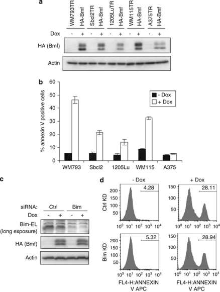
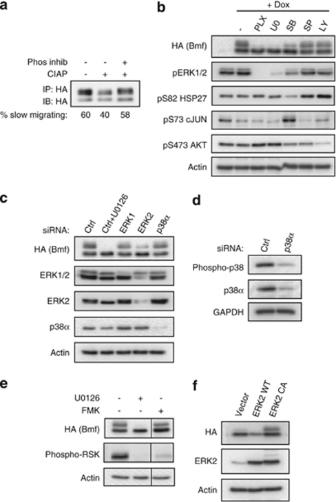
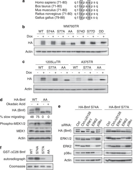
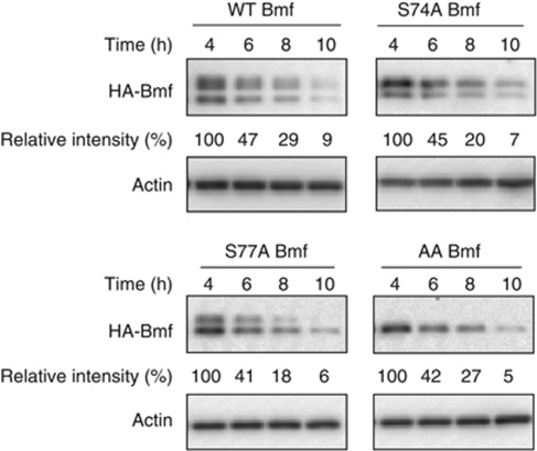
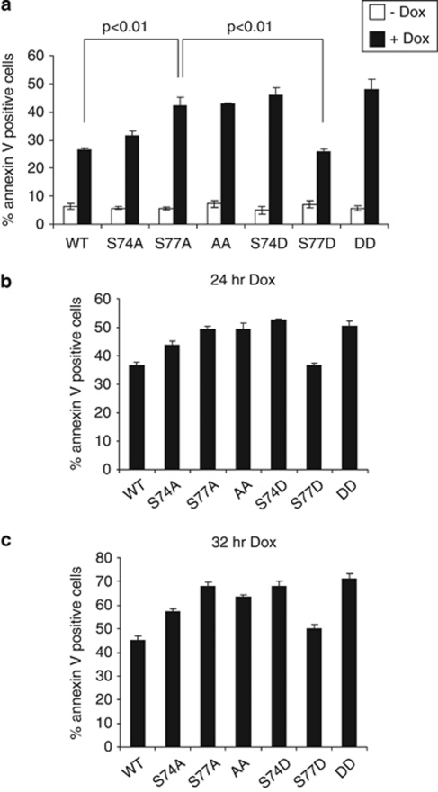
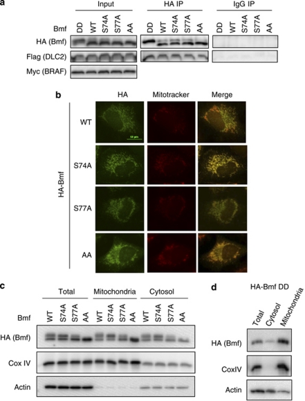
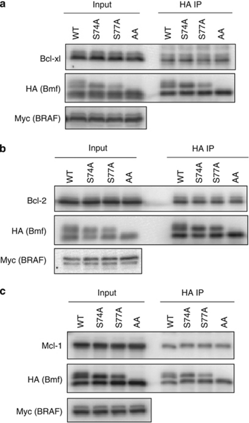
B-cell lymphoma 2 (Bcl-2) homology 3 (BH3)-only proteins represent a class of pro-apoptotic factors that neutralize pro-survival Bcl-2 proteins, and, in some cases, directly activate Bax. The mechanisms of control and the role of BH3-only proteins, such as Bcl-2 like protein 11 extra large and Bad are well studied. By contrast, relatively little is known about the regulation and role of Bcl-2 modifying factor (Bmf). The B-RAF oncogene is mutated in ∼8% of human tumors. We have previously shown that Bmf is upregulated at the transcript level and is required for apoptosis induced by targeting B-RAF signaling in tumor cells harboring mutant B-RAF. In this study, we show that Bmf is regulated at the post-translational level by mutant B-RAF-MEK-ERK2 signaling. Extracellular signal-regulated kinase (ERK2) directly phosphorylates Bmf on serine 74 and serine 77 residues with serine 77 being the predominant site. In addition, serine 77 phosphorylation reduces Bmf pro-apoptotic activity likely through a mechanism independent of altering Bmf localization to the mitochondria and/or interactions with dynein light chain 2 and the pro-survival proteins, B-cell lymphoma extra large, Bcl-2 and Mcl-1. These data identify a novel mode of regulation in Bmf that modulates its pro-apoptotic activity in mutant B-RAF tumor cells.


Figures

Figure 1
Figure 1
Bmf induces apoptosis in melanoma cells independent of Bim-EL. (a) WM793TR/HA-Bmf, Sbcl2TR/HA-Bmf, 1205LuTR/HA-Bmf, WM115TR/HA-Bmf and A375TR/HA-Bmf cells were treated with or without 100 ng/ml doxycycline for 7 h. Cell lysates were analyzed by western blotting using HA-tag and actin (loading control) antibodies. (b) As for a, except that cells were induced with or without 100 ng/ml doxycycline for 48 h. Cells were then stained with annexin-V-APC for flow cytometry analysis. Quantitated are the mean percentages of annexin-V-positive cells from three independent experiments. Error bars: standard deviation. (c) WM793TR/HA-Bmf cells were transfected with control or Bim siRNA for 72 h. Cells were then treated with or without 100 ng/ml doxycycline for additional 24 h before lysis for western blot analysis. (d) As in c except that after 24 h of doxycycline treatment, cells were subject to annexin-V staining and flow cytometry analysis. Representative flow traces from two independent experiments flow traces are shown. x axis, fluorescence intensity; y axis, cell numbers
Figure 2
Figure 2
ERK2 signaling regulates Bmf phosphorylation. (a) A375TR/HA-Bmf WT cells were treated with 100 ng/ml doxycycline overnight and followed by immunoprecipitation using HA-tag antibody. The immunoprecipitates were treated with CIAP or CIAP plus phosphatase inhibitor for 1 h before western blot analysis. Indicated are the percentages of Bmf that is slow migrating, as determined by quantitation of band intensity. (b) WM793TR/HA-Bmf cells were treated with or without 100 ng/ml doxycycline and PLX4720, U0126, SB203580, SP600125, LY294002, as indicated for 7 h. Cell lysates were analyzed by western blotting using the antibodies indicated. Actin serves as a loading control. (c) WM793TR/HA-Bmf cells were transfected with control, ERK1, ERK2 and p38α siRNA. Seventy-two hours post transfection, cells were treated with 100 ng/ml doxycycline and with or without 10 μM U0126 for 7 h before lysis for western blot analysis. (d) Cells were transfected with control non-targeting or p38α-specific siRNAs for 72 h before lysis for western blot analysis using phospho-p38, p38α and GAPDH (loading control) antibodies. (e) WM793TR/HA-Bmf cells were treated with 100 ng/ml doxycycline plus DMSO, 10 μM U0126 or 1 μM FMK for 7 h. Lysates were analyzed by western blotting using antibodies against HA-Tag, phospho-RSK and actin (loading control). (f) HA-Bmf was co-expressed with wild-type ERK2 (ERK2 WT) or constitutive active ERK2 (ERK2 CA) or empty vector in 293FT cells. The effect of ERK2 on Bmf mobility shift was examined by western blot analysis
Figure 3
Figure 3
ERK2 directly phosphorylates residue serine 74 and serine 77 in Bmf. (a) Alignment of Bmf protein sequences surrounding serine 74 and serine 77 among different species. Conserved serine 74 and serine 77 residues are shaded. The amino-acid positions relative to the start codon are shown in parentheses. (b) WM793TR cell lines expressing wild type, S74A, S77A, S74A/S77A (AA), S74D, S77D, S74D/S77D (DD) HA-Bmf were induced with with or without 100 ng/ml doxycycline for 7 h. Cells lysates were analyzed by western blotting using antibodies against the HA-tag and actin. (c) 1205LuTR/HA-Bmf (WT, S77A and AA) cells and A375TR/HA-Bmf (WT, S77A and AA) cells were treated with or without 100 ng/ml doxycycline for 7 h. Cell lysates were analyzed as in b. (d) WM793TR/HA Bmf cells were treated with 100 ng/ml doxycycline for 1.5 h and 1 μM okadaic acid was added for another 30 min. Cells were then lysed for western blot analysis. Indicated are the percentages of Bmf that is slow migrating, as determined by quantitation of band intensity. (e) WM793TR/HA-Bmf S74A and WM793TR/HA-Bmf S77A cells were transfected with control, ERK1, ERK2 and p38α siRNA. Seventy-two hours post transfection, cells were treated with 100 ng/ml doxycycline and with or without 10 μM U0126 for 7 h before lysis for western blot analysis on indicated proteins. (f) Activated ERK2 was incubated with bacterially expressed GST-Bmf WT, GST-Bmf S74A, GST-Bmf S77A and GST-Bmf AA in the presence of [γ-32P]ATP. Phosphorylation of GST-Bmf variants was examined by SDS-PAGE followed by autoradiography. Coomassie staining of the same gel is shown below
Figure 4
Figure 4
Serine 74 and serine 77 phosphorylation does not affect Bmf protein stability. WM793TR/HA-Bmf cell lines (WT, S74A, S77A and AA) were individually treated with 100 ng/ml doxycycline. After 1 h, cells were washed in fresh medium and cultured with medium containing 1 μg/ml actinomycin D but without doxycycline for additional 10 h. During this time course, cell samples were removed after 4, 6, 8 and 10 h for western blot analysis using HA-tag and actin antibodies. The relative intensities of HA-Bmf bands (normalized against actin) were shown below the HA-Bmf blot
Figure 5
Figure 5
Phosphorylation on serine 77 reduces apoptotic activity of Bmf. (a) WM793TR cell lines engineered to inducibly express HA-Bmf variants (WT, S74A, S77A, AA, S74D, S77D and DD) were treated with or without 100 ng/ml doxycycline for 16 h. Cells were stained with annexin-V-APC and analyzed by flow cytometry. Quantitated are the mean percentages of annexin-V-positive cells from three independent experiments. Error bars: standard deviation. (b) As in a except that cells were treated with 100 ng/ml doxycycline for 24 h. (c) As in a except that cells were treated with 100 ng/ml doxycycline for 32 h
Figure 6
Figure 6
Phosphorylation of serine 74 and serine 77 does not alter Bmf association with DLC2 and localization to the mitochondria. (a) HA-Bmf variants (DD, WT, S74A, S77A and AA) were co-expressed with FLAG-tagged DLC2 and Myc-tagged B-RAFV600E in 293FT cells for 24 h. Immunoprecipitation was performed using normal Rabbit IgG or antibody against the HA epitope tag. The presence of HA-Bmf and FLAG-DLC2 in immunoprecipitates and levels of transgene expression in cell lysates (input) was examined by western blot analysis using antibodies against HA and FLAG epitope tags. (b) WM793TR/HA-Bmf WT, S74A, S77A and AA cells were simultaneously treated with 100 ng/ml doxycycline (1 h) and 200 nM mitotracker Red CMXRos (30 min). Cells were then fixed for immunofluoresence analysis. HA-tag antibody was used to detect HA-Bmf (green, left panel). Mitochondria were highlighted by Red CMXRos dye (red, middle panel). The merged images of HA-Bmf and mitochondria staining are shown on the right. (c) Mitochondria fractionation was performed on doxycycline-treated WM793TR/HA-Bmf WT, S74A, S77A and AA cells. Expression of HA-Bmf in total lysates, mitochondria and cytosol fraction was detected by western blot. Actin and Cox IV were used as controls. (d) As in c except WM793TR/HA-Bmf WT DD cells were used
Figure 7
Figure 7
Phosphorylation of serine 74 and serine 77 does not alter Bmf association with pro-survival proteins. HA-Bmf variants (WT, S74A, S77A and AA) were co-expressed in 293 FT cells with Myc-tagged B-RAFV600E and (a) Bcl-xL, (b) Bcl-2 or (c) Mcl-1 for 24 h. Immunoprecipitation was performed using antibody against the HA epitope tag. The presence of Bcl-xL, Bcl-2 and Mcl-1 in immunoprecipitates (HA IP) and levels of transgene expression in cell lysates (input) was examined by western blot analysis

Similar articles

Cited by

References

    1. Merino D, Giam M, Hughes PD, Siggs OM, Heger K, O'Reilly LA, et al. The role of BH3-only protein Bim extends beyond inhibiting Bcl-2-like prosurvival proteins. J Cell Biol. 2009;186:355–362. - PMC - PubMed
    1. Certo M, Del Gaizo Moore V, Nishino M, Wei G, Korsmeyer S, Armstrong SA, et al. Mitochondria primed by death signals determine cellular addiction to antiapoptotic BCL-2 family members. Cancer Cell. 2006;9:351–365. - PubMed
    1. Du H, Wolf J, Schafer B, Moldoveanu T, Chipuk JE, Kuwana T. BH3-domains other than Bim and Bid can directly activate BAX/BAK. J Biol Chem. 2010;286:491–501. - PMC - PubMed
    1. Cheng EH, Wei MC, Weiler S, Flavell RA, Mak TW, Lindsten T, et al. BCL-2, BCL-X(L) sequester BH3 domain-only molecules preventing BAX- and BAK-mediated mitochondrial apoptosis. Mol Cell. 2001;8:705–711. - PubMed
    1. Chipuk JE, Moldoveanu T, Llambi F, Parsons MJ, Green DR. The Bcl-2 family reunion. Mol Cell. 2010;37:299–310. - PMC - PubMed

Publication types

MeSH terms