

A necrotic lung ball caused by co-infection with Candida and Streptococcus pneumoniae
- PMID: 22259251
- PMCID: PMC3259690
- DOI: 10.2147/IDR.S24269
A necrotic lung ball caused by co-infection with Candida and Streptococcus pneumoniae
Abstract
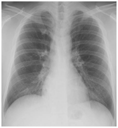
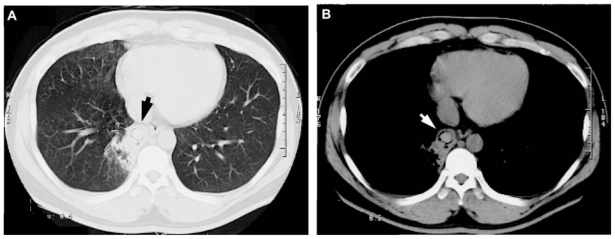
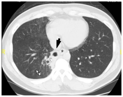
Introduction: A necrotic lung ball is a rare radiological feature that is sometimes seen in cases of pulmonary aspergillosis. This paper reports a rare occurrence of a necrotic lung ball in a young male caused by Candida and Streptococcus pneumoniae.
Case report: A 28-year-old male with pulmonary candidiasis was found to have a lung ball on computed tomography (CT) of the chest. The patient was treated with β-lactams and itraconazole and then fluconazole, which improved his condition (as found on a following chest CT scan) and serum β-D-glucan level. The necrotic lung ball was suspected to have been caused by coinfection with Candida and S. pneumoniae.
Conclusion: A necrotic lung ball can result from infection by Candida and/or S. pneumoniae, indicating that physicians should be aware that patients may still have a fungal infection of the lungs that could result in a lung ball, even when they do not have either Aspergillus antibodies or antigens.
Keywords: Candida; Streptococcus pneumoniae; lung ball; necrotic lung ball.
Figures
Similar articles
-
Pulmonary fungal ball due to Trichophyton successfully managed with oral itraconazole: a case report.J Int Med Res. 2021 Dec;49(12):3000605211066250. doi: 10.1177/03000605211066250. J Int Med Res. 2021. PMID: 34936512 Free PMC article.
-
Treatment of Renal Fungal Ball with Fluconazole Instillation Through a Nephrostomy Tube: Case Report and Literature Review.Am J Case Rep. 2018 Oct 4;19:1179-1183. doi: 10.12659/AJCR.911113. Am J Case Rep. 2018. PMID: 30282963 Free PMC article. Review.
-
[Two cases of pulmonary aspergillosis successfully treated with combinated micafungin and itraconazole therapy].Kansenshogaku Zasshi. 2005 Dec;79(12):951-6. doi: 10.11150/kansenshogakuzasshi1970.79.951. Kansenshogaku Zasshi. 2005. PMID: 16444977 Japanese.
-
Iliopsoas abscess caused by Aspergillus fumigatus complicated by pulmonary aspergillosis.J Infect Chemother. 2012 Aug;18(4):569-75. doi: 10.1007/s10156-011-0339-6. Epub 2011 Nov 15. J Infect Chemother. 2012. PMID: 22080203
-
[Antifungal activity and clinical efficacy of micafungin (funguard)].Nihon Ishinkin Gakkai Zasshi. 2005;46(4):217-22. doi: 10.3314/jjmm.46.217. Nihon Ishinkin Gakkai Zasshi. 2005. PMID: 16282962 Review. Japanese.
Cited by
-
Amino Acid Sensing and Assimilation by the Fungal Pathogen Candida albicans in the Human Host.Pathogens. 2021 Dec 22;11(1):5. doi: 10.3390/pathogens11010005. Pathogens. 2021. PMID: 35055954 Free PMC article. Review.
-
Fungal-bacterial interactions and their relevance to oral health: linking the clinic and the bench.Front Cell Infect Microbiol. 2014 Jul 29;4:101. doi: 10.3389/fcimb.2014.00101. eCollection 2014. Front Cell Infect Microbiol. 2014. PMID: 25120959 Free PMC article. Review.
-
Respiratory syncytial virus infection exacerbates pneumococcal pneumonia via Gas6/Axl-mediated macrophage polarization.J Clin Invest. 2020 Jun 1;130(6):3021-3037. doi: 10.1172/JCI125505. J Clin Invest. 2020. PMID: 32364537 Free PMC article.
-
Innocent until proven guilty: mechanisms and roles of Streptococcus-Candida interactions in oral health and disease.Mol Oral Microbiol. 2014 Jun;29(3):99-116. doi: 10.1111/omi.12049. Mol Oral Microbiol. 2014. PMID: 24877244 Free PMC article. Review.
References
-
- Watanakunakorn C. Acute pulmonary mycetoma due to Candida albicans with complete resolution. J Infect Dis. 1983;148(6):1131. - PubMed
-
- Prats E, Sans J, Valldeperas J, Ferrer JE, Manresa F. Pulmonary mycetoma-like lesion caused by Candida tropicalis. Respir Med. 1995;89(4):303–304. - PubMed
-
- Shelly MA, Poe RH, Kapner LB. Pulmonary mycetoma due to Candida albicans: case report and review. Clin Infect Dis. 1996;22(1):133–135. - PubMed
-
- Abel AT, Parwer S, Sanyal SC. Pulmonary mycetoma probably due to Candida albicans with complete resolution. Respir Med. 1998;92(8):1079–1080. - PubMed
Publication types
LinkOut - more resources
Full Text Sources
