

Fat pad-derived mesenchymal stem cells as a potential source for cell-based adipose tissue repair strategies
- PMID: 22260253
- PMCID: PMC6496625
- DOI: 10.1111/j.1365-2184.2011.00804.x
Fat pad-derived mesenchymal stem cells as a potential source for cell-based adipose tissue repair strategies
Abstract
Background: Mesenchymal stem cells are able to undergo adipogenic differentiation and present a possible alternative cell source for regeneration and replacement of adipose tissue. The human infrapatellar fat pad is a promising source of mesenchymal stem cells with many source advantages over from bone marrow. It is important to determine whether a potential mesenchymal stem-cell exhibits tri-lineage differentiation potential and is able to maintain its proliferation potential and cell-surface characterization on expansion in tissue culture. We have previously shown that mesenchymal stem cells derived from the fat pad can undergo chondrogenic and osteogenic differentiation, and we characterized these cells at early passage. In the study described here, proliferation potential and characterization of fat pad-derived mesenchymal stem cells were assessed at higher passages, and cells were allowed to undergo adipogenic differentiation.
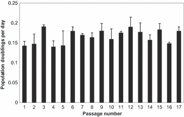
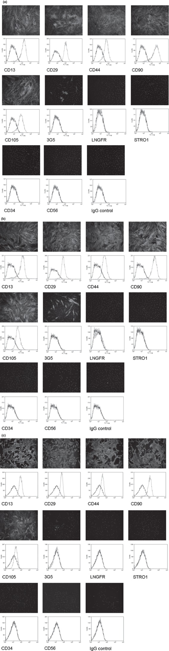
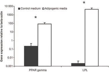
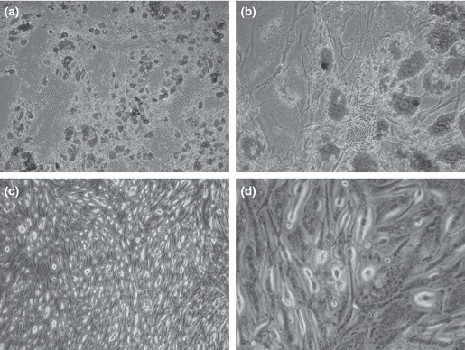
Materials and methods: Infrapatellar fat pad tissue was obtained from six patients undergoing total knee replacement. Cells isolated were expanded to passage 18 and proliferation rates were measured. Passage 10 and 18 cells were characterized for cell-surface epitopes using a range of markers. Passage 2 cells were allowed to undergo differentiation in adipogenic medium.
Results: The cells maintained their population doubling rates up to passage 18. Cells at passage 10 and passage 18 had cell-surface epitope expression similar to other mesenchymal stem cells previously described. By staining it was revealed that they highly expressed CD13, CD29, CD44, CD90 and CD105, and did not express CD34 or CD56, they were also negative for LNGFR and STRO1. 3G5 positive cells were noted in cells from both passages. These fat pad-derived cells had adipogenic differentiation when assessed using gene expression for peroxisome proliferator-activated receptor γ2 and lipoprotein lipase, and oil red O staining.
Discussion: These results indicate that the cells maintained their proliferation rate, and continued expressing mesenchymal stem-cell markers and pericyte marker 3G5 at late passages. These results also show that the cells were capable of adipogenic differentiation and thus could be a promising source for regeneration and replacement of adipose tissue in reconstructive surgery.
© 2012 Blackwell Publishing Ltd.
Figures
References
-
- Patrick CW (2001) Tissue engineering strategies for adipose tissue repair. Anat Rec. 263, 361–366. - PubMed
-
- Beahm EK, Walton RL, Partick CW (2003) Progress in adipose tissue construct development. Clin. Plast. Surg. 30, 547–558. - PubMed
-
- von Heimburg D, Zachariah S, Heschel I, Kühling H, Schoof H, Hafemann B et al. (2001) Human preadipocytes seeded on freeze‐dried collagen scaffolds investigated in vitro and in vivo. Biomaterials 22, 429–438. - PubMed
-
- Patrick CW (2000) Adipose tissue engineering: the future of breast and soft tissue reconstruction following tumor resection. Semin. Surg. Oncol. 19, 302–311. - PubMed
-
- Lee KY, Halberstadt CR, Holder WD, Mooney DJ (2000) Breast reconstruction In: Lanza RP, Langer R, Vacanti J, eds. Principles of Tissue Engineering, pp. 409–423, San Diego, CA: Academic.
MeSH terms
Substances
Grants and funding
LinkOut - more resources
Full Text Sources
Research Materials
Miscellaneous
