Review
. 2012 Apr;30(2):107-16.
doi: 10.3109/08977194.2011.649918. Epub 2012 Jan 20.

EGFR ligands exhibit functional differences in models of paracrine and autocrine signaling

Affiliations
Review

EGFR ligands exhibit functional differences in models of paracrine and autocrine signaling

Kristy J Wilson et al. Growth Factors. 2012 Apr.

Abstract

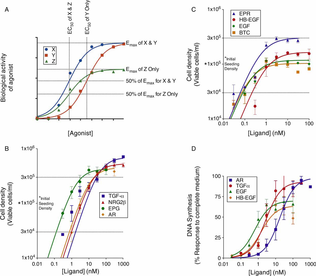
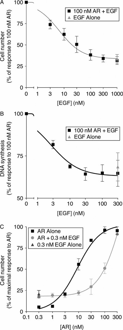
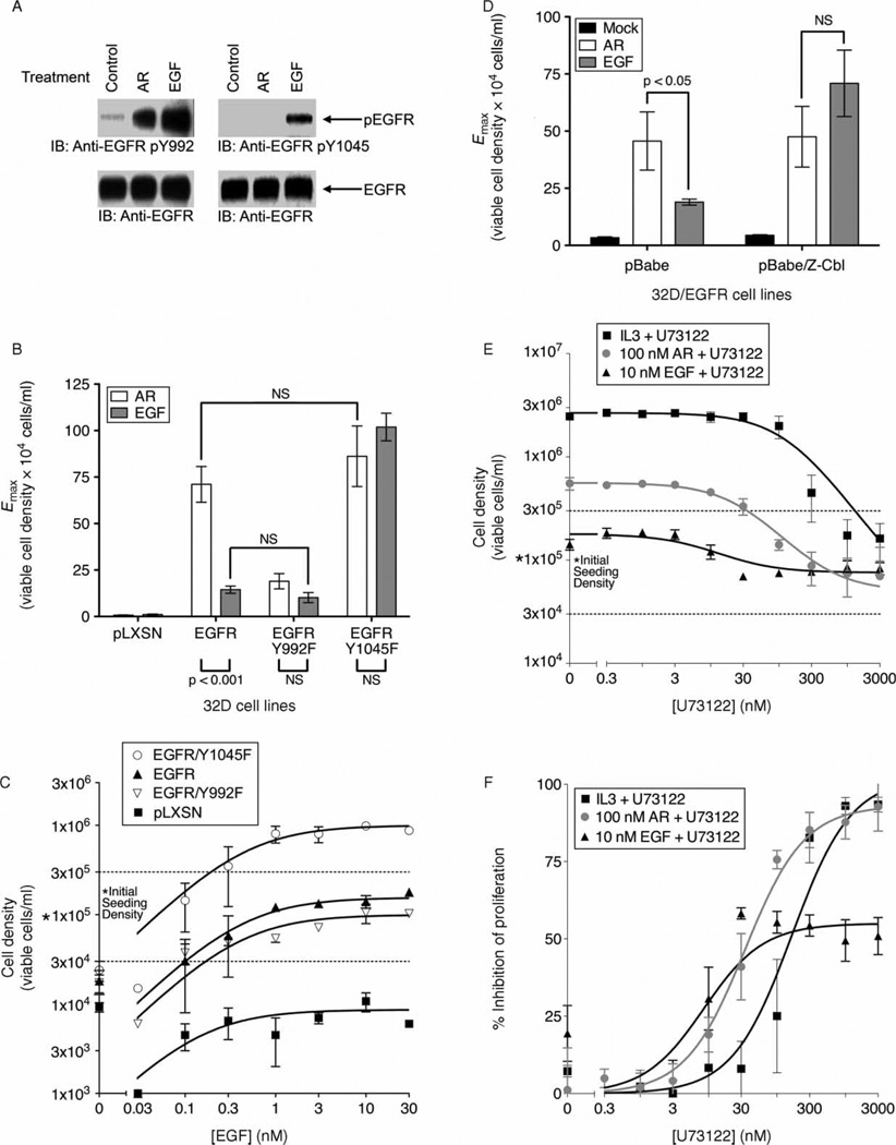
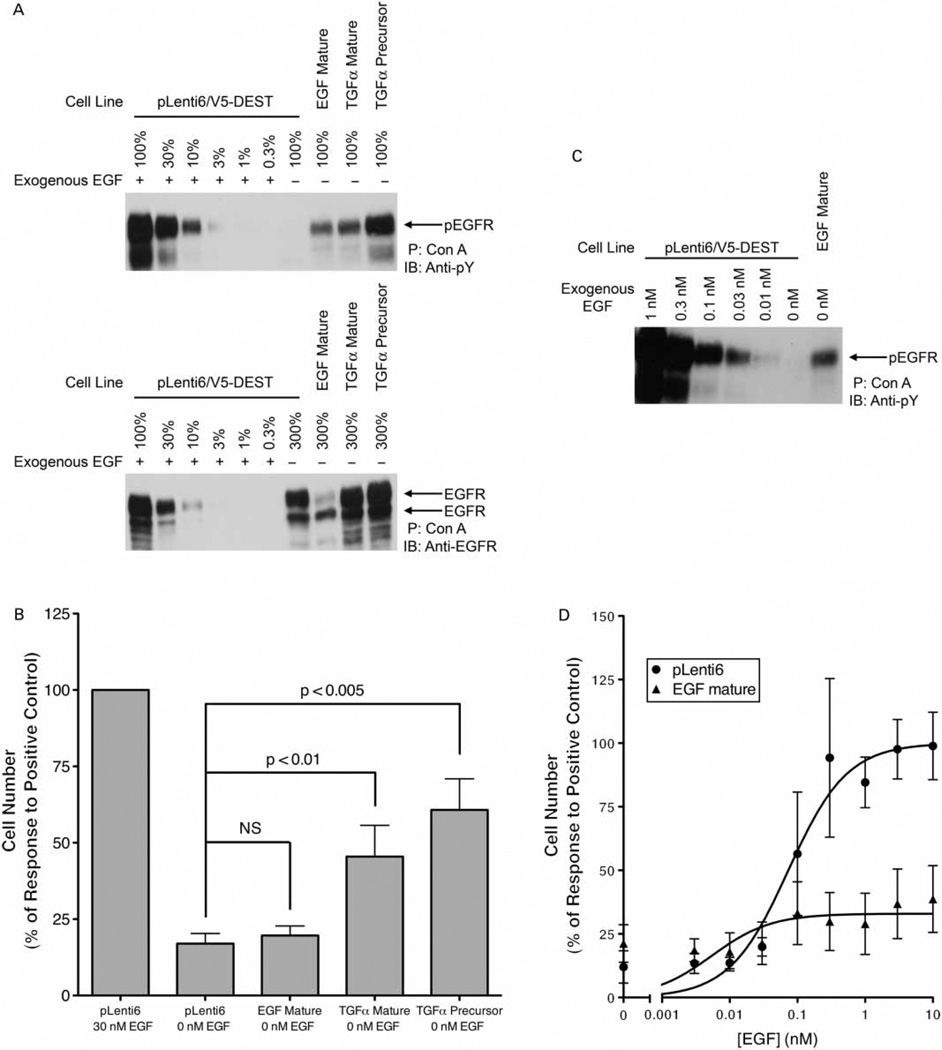
Epidermal growth factor (EGF) family peptides are ligands for the EGF receptor (EGFR). Here, we elucidate functional differences among EGFR ligands and mechanisms underlying these distinctions. In 32D/EGFR myeloid and MCF10A breast cells, soluble amphiregulin (AR), transforming growth factor alpha (TGFα), neuregulin 2 beta, and epigen stimulate greater EGFR coupling to cell proliferation and DNA synthesis than do EGF, betacellulin, heparin-binding EGF-like growth factor, and epiregulin. EGF competitively antagonizes AR, indicating that its functional differences reflect dissimilar intrinsic activity at EGFR. EGF stimulates much greater phosphorylation of EGFR Tyr1045 than does AR. Moreover, the EGFR Y1045F mutation and z-cbl dominant-negative mutant of the c-cbl ubiquitin ligase potentiate the effect of EGF but not of AR. Both EGF and AR stimulate phosphorylation of EGFR Tyr992. However, the EGFR Y992F mutation and phospholipase C gamma inhibitor U73122 reduce the effect of AR much more than that of EGF. Expression of TGFα in 32D/EGFR cells causes greater EGFR coupling to cell proliferation than does expression of EGF. Moreover, expression of EGF in 32D/EGFR cells causes these cells to be largely refractory to stimulation with soluble EGF. Thus, EGFR ligands are functionally distinct in models of paracrine and autocrine signaling and EGFR coupling to biological responses may be specified by competition among functionally distinct EGFR ligands.


Conflict of interest statement

Declaration of Interest: The authors report no conflicts of interest. The authors alone are responsible for the content and writing of the paper.

Figures

Figure 1
Figure 1
EGFR agonists differentially stimulate EGFR coupling in 32D/EGFR cells and MCF10A cells. Data in panels B, C, and D are compiled from at least three independent assays. Error bars indicate the standard error of the means. (A) The simulated agonist dose–response curves depicted here illustrate fundamental principals of pharmacology. Saturating concentrations of agonists X and Y yield the same degree of biological response (Emax). Thus, the concentration- and affinity-independent activities (efficacies or intrinsic activities) of these ligands are identical. Note that the efficacy/intrinsic activity of Z is lower than the efficacies/intrinsic activities of X and Y. The concentration of an agonist that is required to elicit a half-maximal biological response to that agonist (50% of Emax) is termed the EC50. Therefore, the EC50 of X is less than the EC50 of Y and X is a more potent agonist than Y. If X and Y share the same receptor, in many cases X possesses higher affinity for the receptor than does Y. Despite the fact that X possesses greater efficacy/intrinsic activity than does Z, these two agonists possess identical potencies. (B and C) 32D/EGFR cells were treated with increasing concentrations of the indicated EGFR agonists. IL3 independence was evaluated; ligand potency (EC50) and efficacy (Emax) are provided in Table I. (D) DNA synthesis in MCF10A cells was determined following treatment with increasing concentrations of the indicated EGFR agonists. DNA synthesis is expressed as a percentage of that stimulated by complete medium. Ligand potency (EC50) and efficacy (Emax) are provided in Table I.
Figure 2
Figure 2
EGF competitively antagonizes AR stimulation of EGFR coupling to cell proliferation and DNA synthesis. Each data point represents the mean value calculated from at least three independent experiments. Error bars represent the standard error of these means. (A) 32D/EGFR cells were treated with 100 nM AR and/or increasing concentrations of EGF. IL3 independence was determined and is expressed as a percentage of the response to 100 nM AR. (B) DNA synthesis in MCF10A cells was determined following treatment with 100 nM AR and/or increasing concentrations of EGF. DNA synthesis is expressed as a percentage of that stimulated by 100 nM AR. (C) 32D/EGFR cells were treated with increasing concentrations of AR in the presence or absence of 0.3 nM EGF. As a control, cells were treated with 0.3 nM EGF alone. IL3 independence was determined and is expressed as a percentage of the maximal response to AR.
Figure 3
Figure 3
Phosphorylation of EGFR Tyr992 and Tyr1045 specifies EGFR coupling in response to stimulation with EGF or AR. (A) 32D/EGFR cells were stimulated with 10 nM EGF, 100 nM AR, or PBS (diluent control). Immunoblotting was used to assess phosphorylation atTyr992 of EGFR (upper left panel), phosphorylation atTyr1045 of EGFR (upper right panel), and EGFR expression (lower panels). The position of phospho-EGFR (pEGFR) or EGFR on the blots is indicated. Blots are representative of three independent experiments. (B and C) IL3 independence in 32D/LXSN (vector control), 32D/LXSN-EGFR, 32D/LXSN-EGFR/Y992F, and 32D/LXSN-EGFR/Y1045F cells was determined following the treatments described below. (B) Cells were treated with 100 nM AR or 10 nM EGF. Data are averages of at least three independent experiments. Error bars indicate the standard error of the means. A two-way ANOVA with a Bonferroni post-test was used to evaluate statistical significance (p value is indicated) or insignificance (NS). (C) Cells were treated with increasing concentrations of EGF. Data points are averaged from multiple independent experiments. Error bars indicate the standard error of the means. (D) IL3 independence in 32D/LXSN-EGFR/pBabe (vector control) and 32D/LXSN-EGFR/pBabe-z-cbl cells was determined following treatment with 100 nM AR or 10 nM EGF. Data are averages of at least three independent experiments. Error bars indicate the standard error of the means. A two-way ANOVA with a Bonferroni post-test was used to evaluate statistical significance (p value is indicated) or insignificance (NS). (E and F) 32D/EGFR cells were treated with IL3, 10 nM EGF, or 100 nM AR. In addition, cells were treated with increasing concentrations of the PLCγ inhibitor U73122 (Tocris) or DMSO (vehicle control). Data points are averaged from at least three independent experiments. Error bars indicate the standard error of the means. (E) Cell density is plotted as a function of U73122 concentration. (F) Data from the preceding panel are used to plot percent inhibition (relative to maximal stimulation) as a function of U73122 concentration.
Figure 4
Figure 4
Autocrine expression of EGF and TGFα differentially stimulates EGFR coupling. (A) EGFR was precipitated from 32D/EGFR/EGF mature, 32D/EGFR/TGFα mature, and 32D/EGFR/TGFα precursor cell lines. Immunoblotting was used to assess tyrosine phosphorylation (upper panel) and EGFR expression (lower panel). The position of pEGFR or EGFR on the blots is indicated. Increasing amounts of an EGFR precipitate generated from the 32D/EGFR/pLenti6/V5-DEST vector control cell line following stimulation with 30 nM EGF were used to compare EGFR expression and phosphorylation in the experimental cell lines. The figure shown is representative of four independent trials. (B) IL3 independence of 32D/EGFR/EGF mature, 32D/EGFR/TGFα mature, and/or 32D/EGFR/TGFα precursor cell lines was determined. The 32D/EGFR/pLenti6/V5-DEST vector control cell line treated with 30 nM EGF served as the positive control. IL3 independence is expressed as a percentage of this control. The data are averages of at least seven independent experiments; error bars indicate the standard error of the means. A one-way ANOVA was used to evaluate whether differences in viable cell density are statistically significant (p value is indicated) or insignificant (NS). (C) The 32D/EGFR/EGF mature cell line was mock stimulated using diluent (PBS). As a control, the 32D/EGFR/pLenti6/V5-DEST vector control cell line was treated with increasing concentrations of EGF as indicated. EGFR tyrosine phosphorylation was assessed by immunoblotting. The figure shown is representative of five independent experiments. (D) IL3 independence in 32D/EGFR/EGF mature and 32D/EGFR/pLenti6/V5-DEST vector control cell lines was determined following treatment with increasing concentrations of EGF. The data are compiled from seven independent experiments. For each data point, the error bar indicates the standard error of the mean value. EGF potency (EC50) and efficacy (Emax) in each cell line are provided in Table II.

Similar articles

Cited by

References

    1. Baldys A, Gooz M, Morinelli TA, Lee MH, Raymond JR, Jr, Luttrell LM, Raymond JR., Sr Essential role of c-Cbl in amphiregulin-induced recycling and signaling of the endogenous epidermal growth factor receptor. Biochemistry. 2009;48:1462–1473. - PMC - PubMed
    1. Baulida J, Kraus MH, Alimandi M, Di Fiore PP, Carpenter G. All ErbB receptors other than the epidermal growth factor receptor are endocytosis impaired. J Biol Chem. 1996;271:5251–5257. - PubMed
    1. Citri A, Yarden Y. EGF-ERBB signalling: Towards the systems level. Nat Rev Mol Cell Biol. 2006;7:505–516. - PubMed
    1. Duan L, Miura Y, Dimri M, Majumder B, Dodge IL, Reddi AL, Ghosh A, Fernandes N, Zhou P, Mullane-Robinson K, Rao N, Donoghue S, Rogers RA, Bowtell D, Naramura M, Gu H, Band V, Band H. Cbl-mediated ubiquitinylation is required for lysosomal sorting of epidermal growth factor receptor but is dispensable for endocytosis. J Biol Chem. 2003;278:28950–28960. - PubMed
    1. Eden ER, Huang F, Sorkin A, Futter CE. The role of EGF receptor ubiquitination in regulating its intracellular traffic. Traffic. 2011 in press. [Epub ahead of print] - PMC - PubMed

Publication types