. 2012 Sep;16(9):2150-60.
doi: 10.1111/j.1582-4934.2012.01523.x.

MicroRNA-24 regulates cardiac fibrosis after myocardial infarction

Affiliations

MicroRNA-24 regulates cardiac fibrosis after myocardial infarction

Jue Wang et al. J Cell Mol Med. 2012 Sep.

Abstract

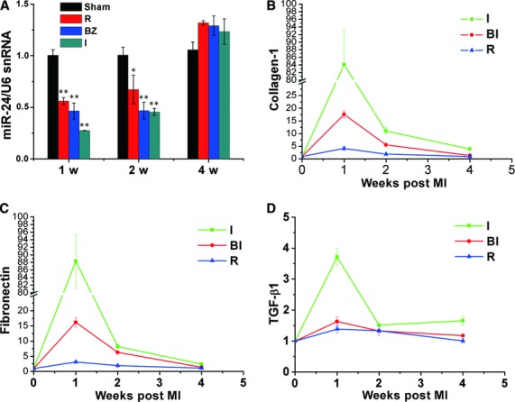
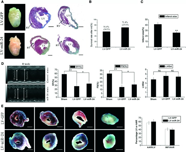
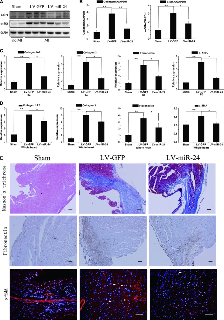
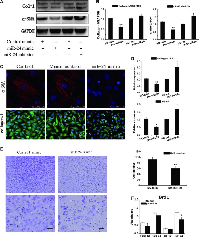
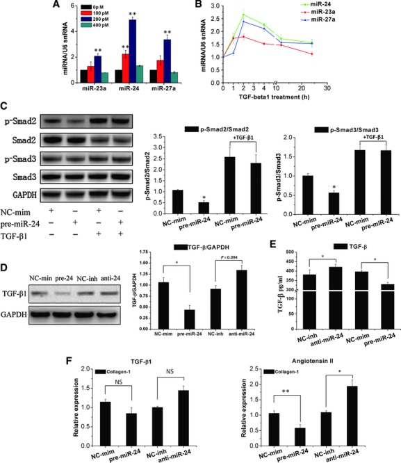
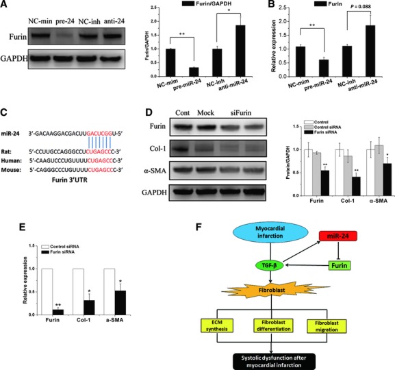
Cardiac fibrosis after myocardial infarction (MI) has been identified as a key factor in the development of heart failure. Although dysregulation of microRNA (miRNA) is involved in various pathophysiological processes in the heart, the role of miRNA in fibrosis regulation after MI is not clear. Previously we observed the correlation between fibrosis and the miR-24 expression in hypertrophic hearts, herein we assessed how miR-24 regulates fibrosis after MI. Using qRT-PCR, we showed that miR-24 was down-regulated in the MI heart; the change in miR-24 expression was closely related to extracellular matrix (ECM) remodelling. In vivo, miR-24 could improve heart function and attenuate fibrosis in the infarct border zone of the heart two weeks after MI through intramyocardial injection of Lentiviruses. Moreover, in vitro experiments suggested that up-regulation of miR-24 by synthetic miR-24 precursors could reduce fibrosis and also decrease the differentiation and migration of cardiac fibroblasts (CFs). TGF-β (a pathological mediator of fibrotic disease) increased miR-24 expression, overexpression of miR-24 reduced TGF-β secretion and Smad2/3 phosphorylation in CFs. By performing microarray analyses and bioinformatics analyses, we found furin to be a potential target for miR-24 in fibrosis (furin is a protease which controls latent TGF-β activation processing). Finally, we demonstrated that protein and mRNA levels of furin were regulated by miR-24 in CFs. These findings suggest that miR-24 has a critical role in CF function and cardiac fibrosis after MI through a furin-TGF-β pathway. Thus, miR-24 may be used as a target for treatment of MI and other fibrotic heart diseases.


Figures

Fig 1
Fig 1
MiR-24 is down-regulated in different areas of the heart after MI. (A) miRNAs were isolated in different areas of the infracted heart and sham-opened hearts 1, 2, and 4 weeks after MI. qRT-PCR was done to determine levels of miR-24 in MI areas and in control groups. Compared with sham-operated hearts, miR-24 expression was significantly decreased in MI areas. (B–D) qRT-PCR was done to determine expression of collagen-1 (B), fibronectin (C), and TGF-β1 (D) in the same samples as (A) (I: infarct zone; BZ: border zone; R: remote zone; n = 3 for each time-point; mean ± S.E.M. *P < 0.05, **P < 0.01 compared with sham groups).
Fig 2
Fig 2
Lentivirus-mediated miR-24 transfer in vivo results in reduced scar formation after MI. (A) Whole images of representative hearts two weeks after MI and Masson's trichrome staining of sections from two representative hearts. miR-24 reduced scar size and built a more compact region in the infarct zone (LV: left ventricle; RV: right ventricle). (B) The number of living and dead mice were counted two weeks after MI: there was no significant difference between LV-miR-24- and LV-GFP-infected groups. (LV-GFP: lentivirus-GFP; LV-miR-24: lentivirus-miR-24). (C) Quantification of infarct size as a percentage of epicardial and endocardial circumference occupied by the infarct on all heart slices of the left ventricle 2 weeks after MI (n = 6); measurements were repeated three times (technical triplicates). MiR-24 transfection resulted in 36.9% reduction in scar size. (D) M-mode echocardiography of representative hearts two weeks after MI. Ejection fraction (EF), and fractional shortening (FS) were measured by using high-resolution echocardiography two weeks after MI. *P < 0.05, **P < 0.01 (n = 9). miR-24 could improve heart function after MI. (E) Representative Evans blue/TTC stains on four continuous slices of left ventricle 24 hrs after MI of control mimic or miR-24 transfections. Blinded quantification of AAR size and infarct size (n = 3). Measurements were repeated thrice (technical triplicates). Bars: (A, E) 1 mm. There was no significant difference between the two groups.
Fig 3
Fig 3
MiR-24 regulates myocardial fibrosis 2 weeks after MI (A, B) Mice receiving control lentiviral vector-GFP (LV-GFP) or lentiviral vector-miR-24 (LV-miR-24) were killed 2 weeks after MI. Protein levels of Col-1, α-SMA and GAPDH in the border zone were determined by western blotting, and densitometric quantification of protein expression (n = 6). (C, D) qRT-PCR was done to determine mRNA levels of Col-1A2, Col-3, fibronectin, and α-SMA in the border zone (C) or whole heart (D) of MI mice. Mean ± S.E.M., *P < 0.05, **P < 0.01 (n = 6). (E) Experiments were done as described in (B). Masson's trichrome blue was used to stain fibrosis, fibronectin was stained to detect ECM remodelling; bars: 100 μm; and α-SMA was stained to detect CF differentiation (arrows denote SMA-positive spindle-shaped myofibroblasts); bars: 50 μm.
Fig 4
Fig 4
MiR-24 down-regulates CF fibrosis through regulation of the differentiation, and migration of CFs. (A, B) CFs were transfected with a pre-miR-24 or anti-miR-24. Expression of collagen-1 and α-SMA was determined by western blotting followed by densitometric quantification of protein expression (n = 6). (NC-mim, negative control mimic; NC-inh, negative control inhibitor). (C) Immunofluorescence showed that up-regulated miR-24 results in a decrease in the expression of α-SMA and collagen-1 protein in CFs compared with control. Bars: 50 μm. (D) Experiments were done as described in (A), and α-SMA and Col-1A2 mRNA levels in CFs were detected by qRT-PCR (n = 6). (E) A transwell migration assay was used to detect the role of miR-24 in CF migration. Representative images of CFs transfected with a control mimic and pre-miR-24 mimic migrated towards 1% serum media. Quantitative analyses demonstrated the difference in CF migration. Mean ± S.E.M., *P < 0.05, **P < 0.01 (n = 3). Bars: 100 μm up two figures, 50 μm down two figures. (F) BrdU incorporation in CFs grown in serum-free (SF) or with 10% foetal bovine serum (FBS) cultured for 24 and 72 hrs after transfection with a pre-miR-24 mimic (n = 3). Notable miR-24 could decrease proliferation of CFs.
Fig 5
Fig 5
MiR-24 is induced by TGF-β1 and regulates the activities of TGF-β1. (A, B) CFs were treated with TGF-β1 at indicated concentrations (100, 200, 400 pM) for 4 hrs, or with 200 pM TGF-β1 for the indicated time. miR-24 cluster levels were determined by qRT-PCR (n = 3). (C) Smad2/3 phosphorylation were detected by western blotting in cultured CFs treated for 24 hrs with a pre-miR-24 mimic and for an additional 24 hrs with TGF-β1 (200 pM), and quantification of Smad2 or Smad3 phosphorylation normalization to total Smad2 and Smad3 (n = 3). (D) Expression of TGF-β protein was detected by western blotting and normalization to GAPDH (n = 3). (E) The TGF-β expression in culture supernatants was measured by ELISA. miR-24 can significantly inhibit TGF-β release compared with control group, and miR-24 inhibitor could induce TGF-β secretion (n = 3). (F) qRT-PCR determined Col-1A2 mRNA expression in CFs transfected with a miR-24 mimic or inhibitor for 24 hrs, and CFs were then treated with TGF-β1 (200 pM) or angiotensin II (100 nM) for an additional 24 hrs. miR-24 can regulate the function of angiotensin II (but not TGF-β1) in CFs. Mean ± S.E.M., *P < 0.05, **P < 0.01 (n = 3).
Fig 6
Fig 6
Target of miR-24 and proposed model of miR-24 function. (A) Expression of furin protein was detected by western blotting and normalization to GAPDH. (B) qRT-PCR revealed furin mRNA expression in CFs transfected with a pre-miR-24 mimic or anti-miR-24 inhibitor. Mean ± S.E.M., *P < 0.05, **P < 0.01 (n = 3). (C) Furin putative miR-24 binding sites (potential complementary residues shown in red). (D) CFs were transfected with siRNA of furin, and expression of furin, Collagen-1 and α-SMA was determined by western blotting and quantitative determination (n = 3). (E) qRT-PCR analysis for Furin, Collagen-1 and α-SMA mRNA expression. Mean ± S.E.M. *P < 0.05. **P < 0.01 compared with control siRNA group (n = 4). (F) Proposed model for the regulation of miR-24 in fibrosis after myocardial infarction.

References

    1. van den Borne SW, Diez J, Blankesteijn WM, et al. Myocardial remodeling after infarction: the role of myofibroblasts. Nat Rev Cardiol. 2010;7:30–7. - PubMed
    1. Zamilpa R, Lindsey ML. Extracellular matrix turnover and signaling during cardiac remodeling following MI: causes and consequences. J Mol Cell Cardiol. 2010;48:558–63. - PMC - PubMed
    1. Tomasek JJ, Gabbiani G, Hinz B, et al. Myofibroblasts and mechano-regulation of connective tissue remodelling. Nat Rev. 2002;3:349–63. - PubMed
    1. Bujak M, Frangogiannis NG. The role of TGF-beta signaling in myocardial infarction and cardiac remodeling. Cardiovasc Res. 2007;74:184–95. - PMC - PubMed
    1. Ambros V. The functions of animal microRNAs. Nature. 2004;431:350–5. - PubMed

Publication types

MeSH terms