

Probing cellular traction forces with magnetic nanowires and microfabricated force sensor arrays
- PMID: 22260885
- PMCID: PMC3376533
- DOI: 10.1088/0957-4484/23/7/075101
Probing cellular traction forces with magnetic nanowires and microfabricated force sensor arrays
Abstract
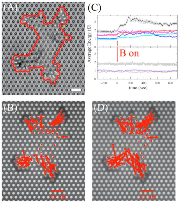
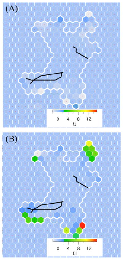
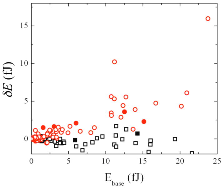
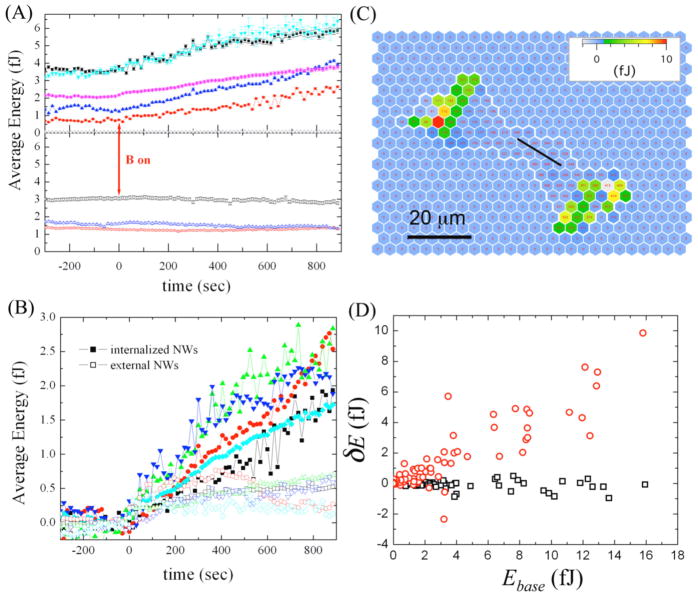
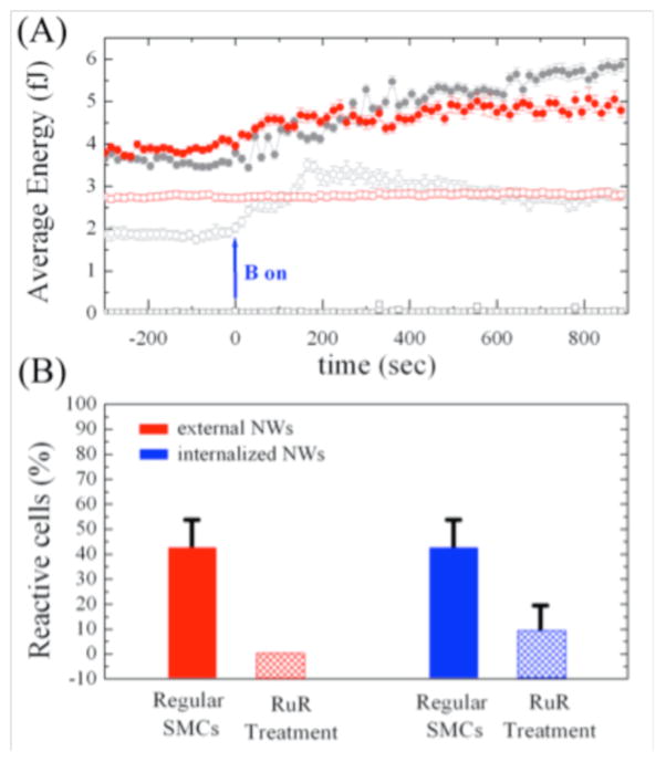
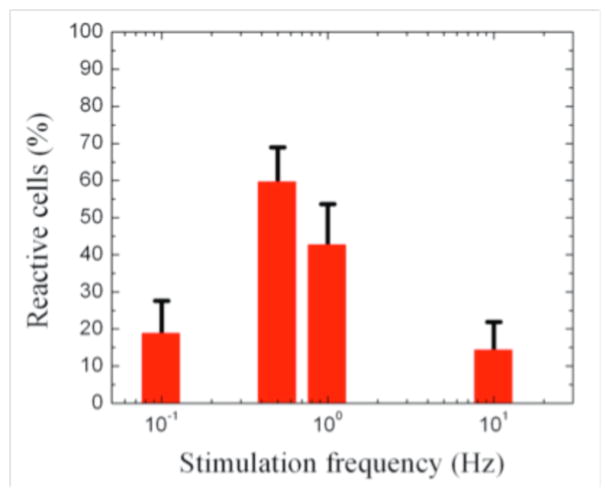
In this paper, the use of magnetic nanowires for the study of cellular response to force is demonstrated. High-aspect ratio Ni rods with diameter 300 nm and lengths up to 20 μm were bound to or internalized by pulmonary artery smooth muscle cells (SMCs) cultured on arrays of flexible micropost force sensors. Forces and torques were applied to the cells by driving the nanowires with AC magnetic fields in the frequency range 0.1-10 Hz, and the changes in cellular contractile forces were recorded with the microposts. These local stimulations yield global force reinforcement of the cells' traction forces, but this contractile reinforcement can be effectively suppressed upon addition of a calcium channel blocker, ruthenium red, suggesting the role of calcium channels in the mechanical response. The responsiveness of the SMCs to actuation depends on the frequency of the applied stimulation. These results show that the combination of magnetic nanoparticles and micropatterned, flexible substrates can provide new approaches to the study of cellular mechanotransduction.
Figures
Similar articles
-
Review of cellular mechanotransduction on micropost substrates.Med Biol Eng Comput. 2016 Mar;54(2-3):249-71. doi: 10.1007/s11517-015-1343-2. Epub 2015 Aug 6. Med Biol Eng Comput. 2016. PMID: 26245253 Review.
-
A novel patterned magnetic micropillar array substrate for analysis of cellular mechanical responses.J Biomech. 2017 Dec 8;65:194-202. doi: 10.1016/j.jbiomech.2017.10.017. Epub 2017 Oct 25. J Biomech. 2017. PMID: 29126605
-
Magnetic microposts as an approach to apply forces to living cells.Proc Natl Acad Sci U S A. 2007 Sep 11;104(37):14553-8. doi: 10.1073/pnas.0611613104. Epub 2007 Sep 5. Proc Natl Acad Sci U S A. 2007. PMID: 17804810 Free PMC article.
-
Magnetic microposts for mechanical stimulation of biological cells: fabrication, characterization, and analysis.Rev Sci Instrum. 2008 Apr;79(4):044302. doi: 10.1063/1.2906228. Rev Sci Instrum. 2008. PMID: 18447536 Free PMC article.
-
Microfabricated tissues for investigating traction forces involved in cell migration and tissue morphogenesis.Cell Mol Life Sci. 2017 May;74(10):1819-1834. doi: 10.1007/s00018-016-2439-z. Epub 2016 Dec 22. Cell Mol Life Sci. 2017. PMID: 28008471 Free PMC article. Review.
Cited by
-
Applications, Surface Modification and Functionalization of Nickel Nanorods.Materials (Basel). 2017 Dec 28;11(1):45. doi: 10.3390/ma11010045. Materials (Basel). 2017. PMID: 29283415 Free PMC article. Review.
-
Manipulation of Axonal Outgrowth via Exogenous Low Forces.Int J Mol Sci. 2020 Oct 28;21(21):8009. doi: 10.3390/ijms21218009. Int J Mol Sci. 2020. PMID: 33126477 Free PMC article. Review.
-
Review of cellular mechanotransduction on micropost substrates.Med Biol Eng Comput. 2016 Mar;54(2-3):249-71. doi: 10.1007/s11517-015-1343-2. Epub 2015 Aug 6. Med Biol Eng Comput. 2016. PMID: 26245253 Review.
-
(De)form and Function: Measuring Cellular Forces with Deformable Materials and Deformable Structures.Adv Healthc Mater. 2020 Apr;9(8):e1901454. doi: 10.1002/adhm.201901454. Epub 2020 Jan 17. Adv Healthc Mater. 2020. PMID: 31951099 Free PMC article. Review.
-
A microfabricated magnetic actuation device for mechanical conditioning of arrays of 3D microtissues.Lab Chip. 2015 Jun 7;15(11):2496-503. doi: 10.1039/c4lc01395f. Epub 2015 May 11. Lab Chip. 2015. PMID: 25959132 Free PMC article.
References
Publication types
MeSH terms
Substances
Grants and funding
LinkOut - more resources
Full Text Sources