. 2012 Apr;60(4):301-15.
doi: 10.1369/0022155411436131. Epub 2012 Jan 19.

Expression and cellular localization of inducible nitric oxide synthase in lipopolysaccharide-treated rat kidneys

Affiliations

Expression and cellular localization of inducible nitric oxide synthase in lipopolysaccharide-treated rat kidneys

Jae-Youn Choi et al. J Histochem Cytochem. 2012 Apr.

Abstract

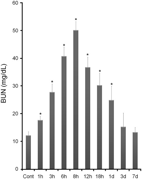
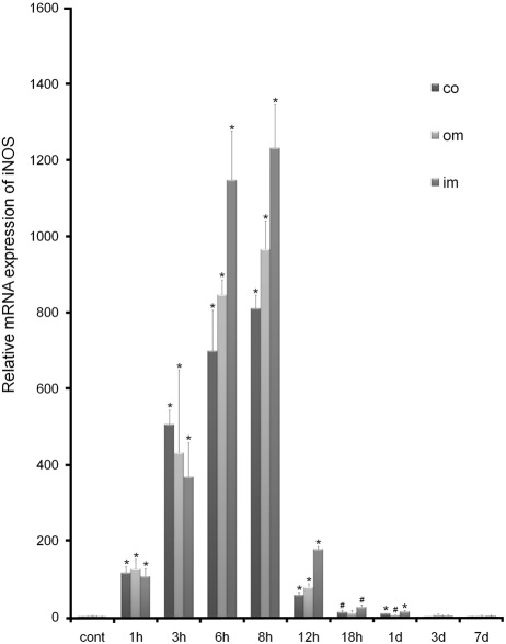
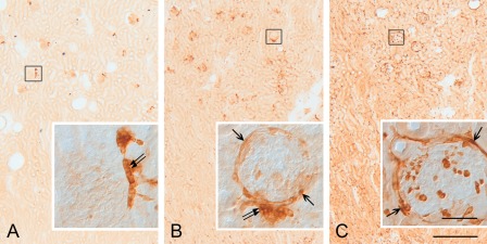
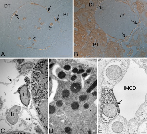
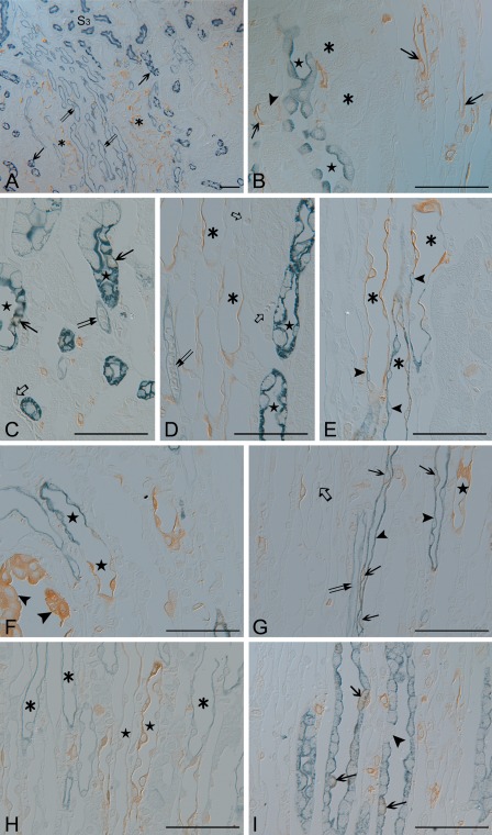
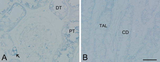
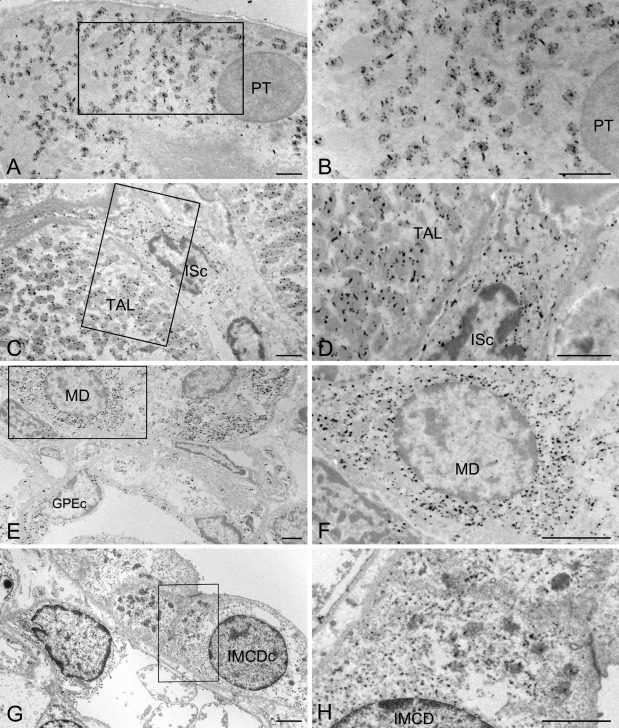
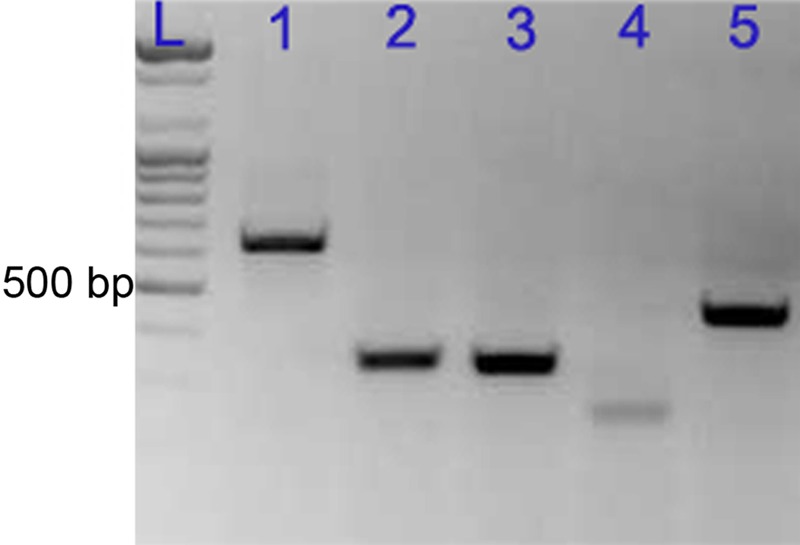
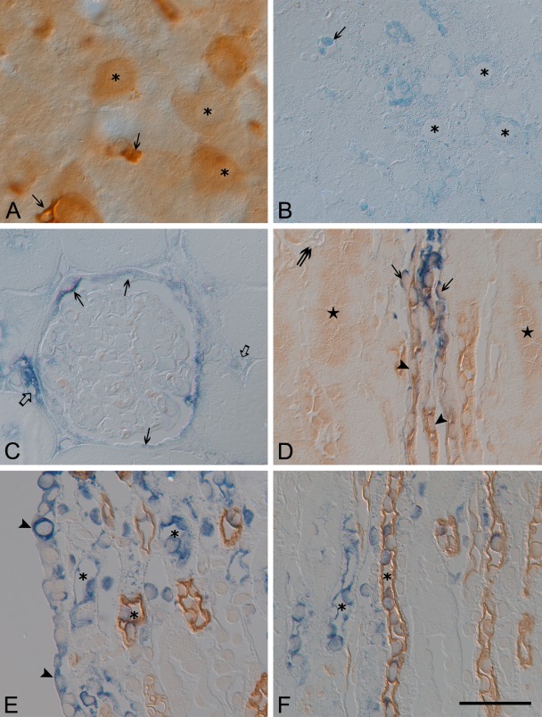
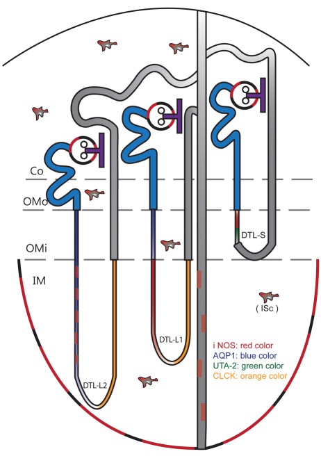
Although inducible nitric oxide synthase (iNOS) is known to play significant roles in the kidney, its renal localization has long been controversial. To resolve this issue, the authors identified iNOS-positive cell types in rat kidneys using double immunohistochemistry and confirmed iNOS positivity using enzyme histochemistry with NADPH-diaphorase (NADPH-d) and in situ RT-PCR. Adult male Sprague-Dawley rats were injected intraperitoneally with lipopolysaccharide (LPS) or saline as a control and sacrificed at various time intervals after injection. Quantitative real-time reverse transcriptase polymerase chain reaction showed that iNOS was not expressed in control kidneys but was induced in LPS-treated kidneys. iNOS immunostaining was strongest 6 to 18 hr after injection and decreased gradually to control levels by day 7. Double immunohistochemistry and NADPH-d revealed that iNOS expression was induced in the interstitial cells, glomerular parietal epithelial cells, the proximal part of the short-looped descending thin limb, the upper and middle papillary parts of the long-looped descending thin limb, some inner medullary collecting duct cells, and almost all calyceal and papillary epithelial cells. The present study determines the precise localization of iNOS in LPS-treated rat kidneys and provides an important morphological basis for examining the roles of iNOS in sepsis-induced acute kidney injury.


Conflict of interest statement

The authors declared no potential conflicts of interest with respect to the authorship and/or publication of this article.

Figures

Figure 1.
Figure 1.
Changes in serum blood urea nitrogen (BUN) levels in control and lipopolysaccharide-treated (LPS-t) rats. *p<0.01 versus control.
Figure 2.
Figure 2.
Real-time PCR analysis of inducible nitric oxide synthase (iNOS) in control and lipopolysaccharide-treated (LPS-t) rat kidneys. The expression level of iNOS was calculated using the comparative threshold cycle method (2–ΔΔCt) with GAPDH as the control gene. The expression level of iNOS in the control was set to 1. co, cortex; im, inner medulla; om, outer medulla. *p<0.01; #p<0.05 versus control.
Figure 3.
Figure 3.
Vibratome sections (50 µm thick) of the renal cortex from the control (A) and 8-hr lipopolysaccharide-treated (LPS-t) rat kidney (B,C) groups immunostained using mouse inducible nitric oxide synthase (iNOS) antibody (610431) (A,B) and rabbit iNOS antibody (610332) purchased from BD Transduction Laboratories (C). For the 610431 antibody immunohistochemistry, no immunostained structure except the macula densa (double arrow in inset of A) was detected in control rat kidneys (A). Note that the immunostaining result using the 610431 antibody is very similar to that using the 610332 antibody except for the macula densa staining (double arrow in inset of B). Arrows in insets of B and C indicate immunostained glomerular parietal epithelial cells. Scale bars = 50 (inset), 500 µm.
Figure 4.
Figure 4.
Light (A,B) and transmission electron (C–E) micrographs for the 8-hr lipopolysaccharide-treated (LPS-t) rat kidney group immunostained using rabbit anti-NOS2 purchased from Santa Cruz Biotechnology. (A) Using a higher dilution (1:1000), proximal (PT) and distal tubules (DT) were completely immunonegative. (B) The PT and DT immunostained with a lower dilution of the antibody (1:100). (A,B) Note the inducible nitric oxide synthase (iNOS)–positive glomerular parietal epithelial cells (arrows), the wandering cells in the glomerular capillary lumen (open arrows), and the iNOS-negative glomerular tufts. (C) An interstitial cell (I2) and PT are densely stained with a lower dilution of the antibody. Note that another interstitial cell (I1) and intertubular vascular endothelia (arrows) are iNOS negative. (D) Magnification of the boxed area in C. Immunoreactivity in the proximal tubule was localized mainly in mitochondria (Mt). (E) In the inner medullary collecting duct (IMCD), an iNOS-positive cell (arrow) is observed. Scale bars = 30 (A, B), 2 (C, E), 0.2 (D) µm.
Figure 5.
Figure 5.
Semithin sections (1.5 µm thick) from the 8-hr lipopolysaccharide-treated (LPS-t) group kidneys double-immunostained for inducible nitric oxide synthase (iNOS) (brown; A–I) and AQP1 (blue; A,C,D,F,G), AQP2 (blue; B,I), CLC-K (blue; H), and UT-A2 (blue; E). (A) iNOS-positive thin tubular structures (asterisks) were observed in the outer and middle layers of the vascular bundle (descending vasa recta; double arrows). The long-looped descending thin limb (DLT) cells (arrows) were located in the interbundle region. (B) Outer medullary collecting duct cells (stars) and the thick ascending limb (asterisks) were iNOS negative. Arrows indicate iNOS positivity in the thin limb, which continues up to the S3 segment of the proximal tubule (arrowhead). (C–E) The short-looped DTL (asterisks), identified by UT-A2 immunolabeling (arrowheads) in the distal portion, was AQP1 negative and iNOS positive. In the long-looped DTL (stars) of the outer medulla, only a few cells expressed iNOS (arrows). The descending (double arrows) and ascending (open arrows) vasa recta were iNOS negative. (F) iNOS-positive epithelia lining the forniceal area (arrowheads). Below this level, AQP1 positivity was lost, but iNOS immunoreactivity was observed in the shorter long-looped DTL (DTL-L1; stars). (G) Intermittent expression of iNOS (arrows) in the AQP1-positive longer long-looped DTL (DTL-L2; arrowheads) at the upper papillary region. In contrast, most cells expressed iNOS in the AQP1-negative DTL-L1 (star). The descending (double arrows) and ascending vasa recta (open arrows) were iNOS negative. (H) In the ascending thin limb (asterisks), no iNOS was detected. In contrast, iNOS was detected in the two types of DTLs (stars). (I) Some inner medullary collecting duct cells (arrows) coexpressed both AQP2 and iNOS. The arrowhead indicates an AQP2-negative intercalated cell. Scale bars = 50 µm.
Figure 6.
Figure 6.
Semithin sections (1.5 µm thick) of NADPH-diaphorase (NADPH-d) enzyme histochemistry for the 8-hr lipopolysaccharide-treated (LPS-t) group kidneys. The reaction particles exhibited punctuated patterns in the cytoplasm of the proximal (PT) and distal (DT) tubules in cortex (A), as well as the collecting ducts (CD) and the thick ascending limb (TAL) in the outer medulla (B). The arrow in A indicates the macula densa. Scale bar = 30 µm.
Figure 7.
Figure 7.
Electron micrographs of NADPH-diaphorase (NADPH-d) enzyme histochemistry for the 8-hr lipopolysaccharide-treated (LPS-t) group kidneys. B, D, F, and H are magnification images of the boxed areas in A, C, E, and G, respectively. In the proximal tubular cell (PT) and thick ascending limb cell (TAL), the reaction products were located exclusively in the mitochondria. In the macular densa (MD), glomerular parietal epithelial cells (GPEc), interstitial cells (ISc), and inner medullary collecting duct cells (IMCDc), the reaction products were diffusely distributed in the cytoplasm. Scale bars = 2 µm.
Figure 8.
Figure 8.
Reverse transcription (RT)–PCR analysis of five inducible nitric oxide synthase (iNOS) primer sets using total RNA from 8-hr lipopolysaccharide-treated (LPS-t) rat kidney. Lanes: L, ladder; 1, 627 bp; 2, 330 bp; 3, 320 bp; 4, 227 bp; 5, 429 bp.
Figure 9.
Figure 9.
Vibratome sections (50 µm thick) from 8-hr lipopolysaccharide-treated (LPS-t) rat liver immunostained for inducible nitric oxide synthase (iNOS) (A), as well as semithin sections (1.5 µm thick) performed with iNOS in situ RT-PCR (blue) from the 8-hr LPS-t rat liver (B) and kidney (C–F). (A, B) Note strong iNOS immunoreactivity and in situ signals in Kupffer cells (arrow). Various intensities of iNOS in situ signals are shown in hepatic cells (asterisk). (C) Note the iNOS-positive glomerular parietal epithelial cells (arrows), interstitial cells (open arrows), and the iNOS-negative glomerular tufts. (D) The short looped-descending thin limb showing iNOS in situ signal was identified using UT-A2 immunolabeling in the distal portion. In comparison to Figure 6E, UT-A antigenicity was much reduced, and nonspecific staining was increased (stars). The vasa recta (double arrow) showed no iNOS in situ signal. (E, F) Note the papilla epithelial cells (arrowheads) with iNOS signal. The long-looped DTL (DTL-L) (asterisk) of the inner medulla is identified by AQP1 positivity (brown) Bar = 40 um.
Figure 10.
Figure 10.
Diagram illustrating the cellular localization of inducible nitric oxide synthase (iNOS) in lipopolysaccharide-treated (LPS-t) rat kidneys. Co, Cotex; DTL-L1, shorter long-looped DTL; DTL-L2, longer long-looped DTL; DTL-S, short-looped DTL; IM, inner medulla; ISc, interstitial cells; OMi, inner stripe of outer medulla; OMo, outer stripe of outer medulla.

Similar articles

Cited by

References

    1. Abdel-latief M, Garbe LA, Koch M, Ruther J. 2008. An epoxide hydrolase involved in the biosynthesis of an insect sex attractant and its use to localize the production site. Proc Natl Acad Sci U S A. 105:8914–8919 - PMC - PubMed
    1. Ahn KY, Mohaupt MG, Madsen KM, Kone BC. 1994. In situ hybridization localization of mRNA encoding inducible nitric oxide synthase in rat kidney. Am J Physiol. 267:F748-F757 - PubMed
    1. Bates TE, Loesch A, Burnstock G, Clark JB. 1996. Mitochondrial nitric oxide synthase: a ubiquitous regulator of oxidative phosphorylation? Biochem Biophys Res Commun. 218:40–44 - PubMed
    1. Buffoli B, Pechánová O, Kojsová S, Andriantsitohaina R, Giugno L, Bianchi R, Rezzani R. 2005. Provinol prevents CsA-induced nephrotoxicity by reducing reactive oxygen species, iNOS, and NF-κB expression. J Histochem Cytochem. 53:1459–1468 - PMC - PubMed
    1. Cha JH, Woo SK, Han KH, Kim YH, Handler JS, Kim J, Kwon HM. 2001. Hydration status affects nuclear distribution of transcription factor tonicity responsive enhancer binding protein in rat kidney. J Am Soc Nephrol. 12:2221–2230 - PubMed

Publication types