. 2012 Jan 18;4(117):117ra7.
doi: 10.1126/scitranslmed.3003008.

Skin effector memory T cells do not recirculate and provide immune protection in alemtuzumab-treated CTCL patients

Affiliations

Skin effector memory T cells do not recirculate and provide immune protection in alemtuzumab-treated CTCL patients

Rachael A Clark et al. Sci Transl Med. .

Abstract

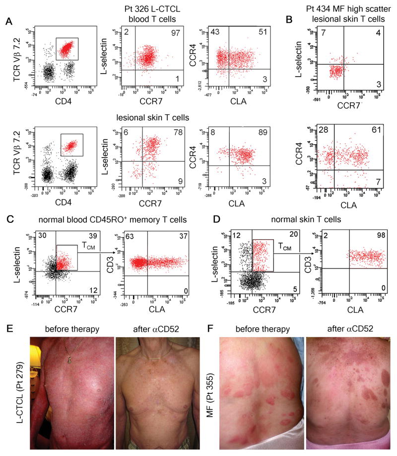
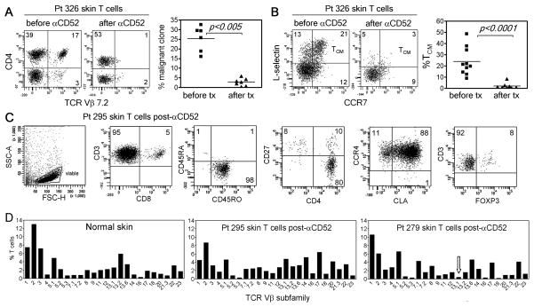
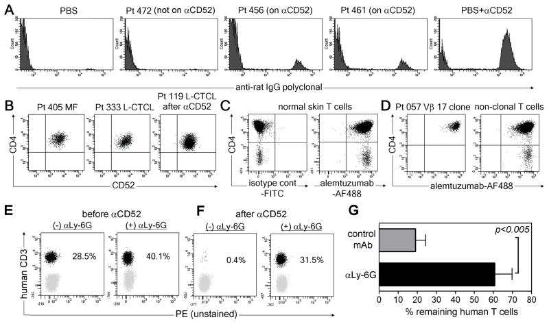
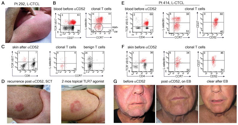
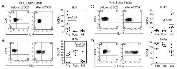
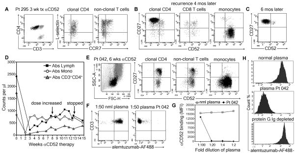
Cutaneous T cell lymphoma (CTCL) is a cancer of skin-homing T cells with variants that include leukemic CTCL (L-CTCL), a malignancy of central memory T cells (T(CM)), and mycosis fungoides (MF), a malignancy of skin resident effector memory T cells (T(EM)). We report that low-dose alemtuzumab (αCD52) effectively treated patients with refractory L-CTCL but not MF. Alemtuzumab depleted all T cells in blood and depleted both benign and malignant T(CM) from skin, but a diverse population of skin resident T(EM) remained in skin after therapy. T cell depletion with alemtuzumab required the presence of neutrophils, a cell type frequent in blood but rare in normal skin. These data suggest that T(CM) were depleted because they recirculate between the blood and the skin, whereas skin resident T(EM) were spared because they are sessile and non-recirculating. After alemtuzumab treatment, skin T cells produced lower amounts of interleukin-4 and higher amounts of interferon-γ. Moreover, there was a marked lack of infections in alemtuzumab-treated L-CTCL patients despite the complete absence of T cells in the blood, suggesting that skin resident T(EM) can protect the skin from pathogens even in the absence of T cell recruitment from the circulation. Together, these data suggest that alemtuzumab may treat refractory L-CTCL without severely compromising the immune response to infection by depleting circulating T(CM) but sparing the skin resident T(EM) that provide local immune protection of the skin.


Conflict of interest statement

Competing interests: C.S.C. has served on an advisory board for Genzyme. R.A.C. served on an expert panel for Biogen Idec. The other authors declare that they have no competing interests.

Figures

Fig. 1
Fig. 1
Low dose alemtuzumab is effective in the treatment of L-CTCL, a malignancy of TCM, but is ineffective in MF, a malignancy of TEM. (A) Clonal malignant T cells isolated from the blood and lesional skin of patients with L-CTCL co-expressed L-selectin and CCR7, a phenotype consistent with TCM. A subset of circulating malignant cells also expressed the skin homing addressins CCR4 and CLA. Malignant T cells co-expressing CCR4 and CLA predominated in skin lesions. A representative patient is shown; expression of CLA and CCR4 was confirmed in 5 additional L-CTCL patients with identifiable T cell clones (Table S1). (B) High side/forward scatter can be used to identify the malignant T cells in CTCL. High scatter cells isolated from the fixed skin lesions of MF expressed skin homing addressins CLA and CCR4 but lacked expression of L-selectin and CCR7, consistent with the claim that MF is a malignancy of skin resident effector memory T cells (TEM). Findings have been replicated in 17 additional MF patients. (C) A subset of circulating TCM in the blood of healthy individuals co-express the skin homing addressin CLA. (D) These CLA-expressing TCM make up approximately 20% of the T cells present in normal human skin. As observed in L-CTCL, only CLA-expressing TCM were found in skin. (E) Alemtuzumab is effective in the treatment of refractory L-CTCL. Patient 279 received six weeks of low dose alemtuzumab (αCD52) and had complete clearance of skin disease and loss of the malignant T cell clone from his blood. (F) In contrast, two MF patients (including the patient shown) experienced no improvement in inflammatory skin lesions with alemtuzumab therapy.
Fig. 2
Fig. 2
Alemtuzumab therapy depletes TCM from both blood and skin but does not affect skin resident TEM. (A) In patient 326, the TCR Vβ7.2-expressing malignant T cell clone was prominent in the skin before treatment but not detectable after alemtuzumab therapy. Similar results in additional patients with identifiable malignant T cell clones are shown. (B) All TCM were depleted from skin by alemtuzumab in patient 326. Studies in additional patients demonstrated that depletion of both benign and malignant TCM from skin was a common feature of alemtuzumab therapy. (C) TEM remaining in the skin of a patient on alemtuzumab therapy are shown. Skin T cells were CD4+ or CD8+ CD45RO+ memory T cells that expressed the skin homing addressins CLA and CCR4. Most CD4+ skin resident T cells were CD27low, a phenotype suggestive of TEM, although a subpopulation of FOXP3+ regulatory T cells was also evident. A representative patient is shown; the TEM phenotype of T cells remaining in skin was confirmed in 6 additional patients (Table S2). (D) The TCR diversity of T cells remaining in skin after alemtuzumab therapy was assessed by flow cytometry and found to be comparable to that of normal skin. The malignant clone in patient 295 was not identified by commercially available TCR Vβ antibodies. The malignant clonotype in patient 279 (TCR Vβ 13.1) is indicated by an arrow. Similar findings were observed in 4 additionalalemtuzumab treated patients.
Fig. 3
Fig. 3
Neutrophils can support alemtuzumab-mediated T cell depletion. (A) Alemtuzumab was detected in the extracellular fluid of skin from patients on alemtuzumab therapy (456, 461) but not in a patient not receiving alemtuzumab (472). Negative (PBS) and positive control (PBS with alemtuzumab; PBS+αCD52) samples are also shown. (B) CD52 is expressed by skin T cells in CTCL patients before and after alemtuzumab therapy. Gates are set based on isotype-matched negative control staining. Representative patients are shown, similar results were observed in seven additional MF patients, 12 additional L-CTCL patients prior to therapy and five additional patients after alemtuzumab therapy (Table S3). (C, D) Alemtuzumab conjugated to AF488 bound to T cells from (C) normal skin and (D) both benign and malignant T cells from CTCL skin lesions. (E–G) NOD/SCID/IL2 receptor γ chainnull mice were engrafted with human PBMC and treated with alemtuzumab in the presence of either the neutrophil depleting Ab αLy-6G ((+) αLy-6G) or control mAb ((−) αLy-6G). The % of human T cells in blood before (E) and after (F) alemtuzumab was determined by flow cytometry. A representative set of mice is shown. (G) Aggregate data (mean+/− SEM) from eight mice treated with control Ab and nine treated with αLy-6G.
Fig. 4
Fig. 4
Alemtuzumab clears central memory (TCM) but not effector memory (TEM) disease. (A) Patient 292 had both diffuse erythema and discrete papules on presentation. (B) Clonal T cells in blood were TCR Vβ17+ with both TCM and TEM phenotypes. (C) After alemtuzumab, skin biopsy showed clearance of TCM from skin but detectable Vβ17+ TEM remained. (D) The patient underwent stem cell transplantation and disease then recurred with isolated skin lesions that responded to topical TLR7 agonist therapy. In a second patient, malignant T cells from the (E) blood and (F) skin were TCR Vβ13.1+ with both TCM and TEM phenotypes.(G) Clinical presentation with diffuse erythema and leonine facies. Well demarcated lesions remained after alemtuzmab. Skin directed electron beam therapy cleared remaining skin disease.
Fig. 5
Fig. 5
Patients treated with alemtuzumab have decreased Th2 cytokine production and enhanced Th1 responses after therapy. (A) IL-4 was produced by greater percentages of T cells from L-CTCL skin lesions as compared to normal skin, and production was decreased after alemtuzumab therapy. Total T cells from L-CTCL skin lesions are shown. Representative histograms and results from multiple donors before (Pre) and after (Post) alemtuzumab are shown, compared to normal skin (NS). (B) Production of IFNγ was enhanced after alemtuzumab therapy. (C) Production of IL-17 was low in untreated L-CTCL patients and did not recover after alemtuzumab therapy. (D) TNFα production trended upward after alemtuzumab therapy but this change was not statistically significant.
Fig. 6
Fig. 6
Down-regulation of CD52 and development of a blocking antibody can lead to alemtuzumab resistance. (A) Malignant T cells from the blood of patient 295 were identifiable by their distinct CD4+CD3low phenotype. At 3 weeks of therapy, both benign (gray) and malignant (black) T cells remained in blood. (B) There was loss of CD52 expression on clonal malignant T cells. (C) Down-regulation of CD52 expression was persistent. (D) Patient 042 showed only transient loss of lymphocytes and monocytes. (E) CD3 T cells and monocytes expressed CD52. (F) Patient plasma blocked binding of alemtuzumab to normal blood T cells. (G) Loss of alemtuzumab binding was titratable and (H) depleted by protein G sepharose.

Comment in

  • T cell memory: Skin-deep memory.
    Bird L. Bird L. Nat Rev Immunol. 2012 Mar 22;12(4):230-1. doi: 10.1038/nri3201. Nat Rev Immunol. 2012. PMID: 22437929 No abstract available.

References

    1. Willemze R, Jaffe ES, Burg G, Cerroni L, Berti E, Swerdlow SH, Ralfkiaer E, Chimenti S, Diaz-Perez JL, Duncan LM, Grange F, Harris NL, Kempf W, Kerl H, Kurrer M, Knobler R, Pimpinelli N, Sander C, Santucci M, Sterry W, Vermeer MH, Wechsler J, Whittaker S, Meijer CJLM. WHO-EORTC classification for cutaneous lymphomas 10.1182/blood-2004-09-3502. Blood. 2005;105:3768. - PubMed
    1. Kim YH, Liu HL, Mraz-Gernhard S, Varghese A, Hoppe RT. Long-term outcome of 525 patients with mycosis fungoides and Sezary syndrome: clinical prognostic factors and risk for disease progression. Archives of dermatology. 2003;139:857. - PubMed
    1. NCCN Clinical Practice Guidelines in Oncology Non-Hodgkin’s Lymphomas Version 3.2009 Vol. 2009 To view the most recent and complete version of the NCCN Guidelines, go online to NCCN.org. (© 2009 National Comprehensive Cancer Network, Inc., 2009)..

    1. Shin J, Monti S, Aires DJ, Duvic M, Golub T, Jones DA, Kupper TS. Lesional gene expression profiling in cutaneous T-cell lymphoma reveals natural clusters associated with disease outcome. Blood. 2007;110:3015. - PMC - PubMed
    1. van Doorn R, van Kester MS, Dijkman R, Vermeer MH, Mulder AA, Szuhai K, Knijnenburg J, Boer JM, Willemze R, Tensen CP. Oncogenomic analysis of mycosis fungoides reveals major differences with Sezary syndrome. Blood. 2009;113:127. - PubMed

Publication types

MeSH terms

LinkOut - more resources