. 2011 Dec 7;101(11):2555-62.
doi: 10.1016/j.bpj.2011.10.043.

The layered structure of coronary adventitia under mechanical load

Affiliations

The layered structure of coronary adventitia under mechanical load

Huan Chen et al. Biophys J. .

Abstract

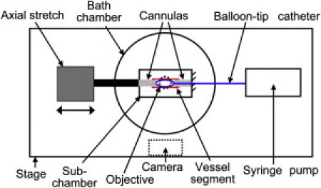
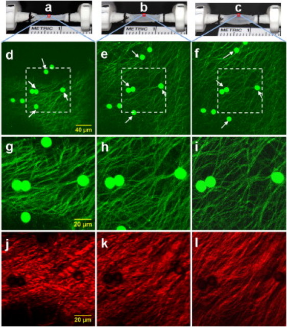
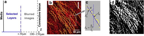
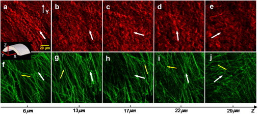
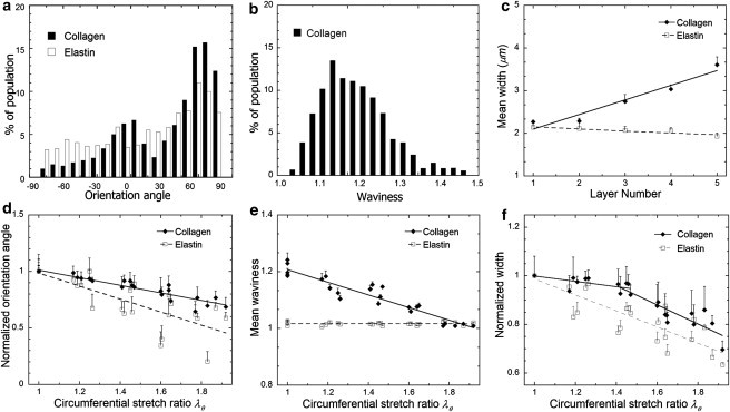
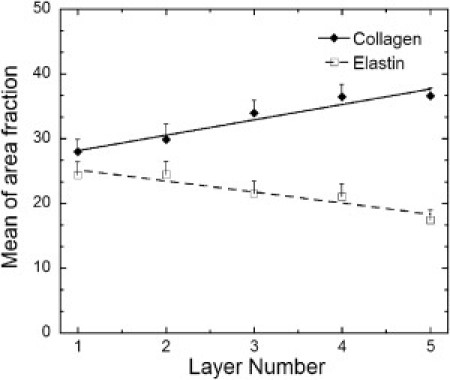
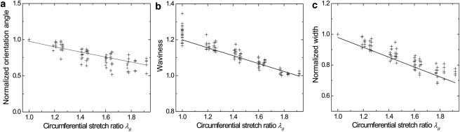
The mechanical loading-deformation relation of elastin and collagen fibril bundles is fundamental to understanding the microstructural properties of tissue. Here, we use multiphoton microscopy to obtain quantitative data of elastin and collagen fiber bundles under in situ loading of coronary adventitia. Simultaneous loading-imaging experiments on unstained fresh coronary adventitia allowed morphometric measurements of collagen and elastin fibril bundles and their individual deformation. Fiber data were analyzed at five different distension loading points (circumferential stretch ratio λ(θ) = 1.0, 1.2, 1.4, 1.6, and 1.8) at a physiological axial stretch ratio of λ(axial) = 1.3. Four fiber geometrical parameters were used to quantify the fibers: orientation angle, waviness, width, and area fraction. The results show that elastin and collagen fibers in inner adventitia form concentric densely packed fiber sheets, and the fiber orientation angle, width, and area fraction vary transmurally. The extent of fiber deformation depends on the initial orientation angle at no-distension state (λ(θ) = 1.0 and λ(axial) = 1.3). At higher distension loading, the orientation angle and waviness of fibers decrease linearly, but the width of collagen fiber is relatively constant at λ(θ) = 1.0-1.4 and then decrease linearly for λ(θ) ≥ 1.4. A decrease of the relative dispersion (SD/mean) of collagen fiber waviness suggests a heterogeneous mechanical response to loads. This study provides fundamental microstructural data for coronary artery biomechanics and we consider it seminal for structural models.


Figures

Figure 1
Figure 1
Schematic diagram of the experimental setup.
Figure 2
Figure 2
MPM images of collagen and elastin fibers under different mechanical loading. Fluorescent microspheres were used to track the scan area and deformation of individual fibers. (ac) Photos of the specimen with the scaling marker in subchamber were taken to record the specimen diameters at (a) no-distension state λθ = 1.0; (b) λθ = 1.63; and (c) λθ = 1.92, respectively. (df) TPEF images with lower magnification were captured first to track the scan area at corresponding loads. (gi) TPEF images (for elastin fiber) with appropriate magnification and resolution were collected at corresponding loads. (jl) SHG images (for collagen fiber) were collected simultaneously with TPEF images.
Figure 3
Figure 3
(a) Selected fiber layers were within ≈30 μm depth of inner adventitia. (b) Center-lines of fibers on a MPM image (arrow indicates the circumferential direction). (c) The orientation angle θ and the waviness λ0 for a single fiber. (d) Threshold of MPM image to a binary image for measurement of the area fraction of fibers.
Figure 4
Figure 4
Both collagen and elastin fibers in inner adventitia form layered structures. Images ae are five collagen layers at different depth Z, and images fj are five elastin layers at the exactly same depth. (White arrow) Main orientation of fibers in all layers. (Yellow arrow) Second principle direction for elastin (X, Y, Z: axial, circumferential, and radial directions) in all layers.
Figure 5
Figure 5
Statistical data of microstructure and deformation of collagen and elastin fibers. At no-distention state (λθ = 1.0 with axial stretch ratio λaxial = 1.3): (a) The orientation distribution of fibers of all specimen layers. (b) The bell-shaped distribution of the collagen fiber waviness of all layers. (c) Layer-to-layer heterogeneity of the fiber width. The curve-fitted linear width-layer relationship of collagen is 1.76 + 0.34N (correlation coefficient R = 0.97, N is the layer number), and that of elastin is 2.20–0.05N (R = 0.92). At different higher loadings: (d) Change of mean orientation angle normalized by initial orientation angle at λθ = 1.0, and fitted linear orientation-distension relationship of collagen fibers is 1.35–0.34λθ (R = 0.94), and that of elastin fibers is 1.56–0.58λθ (R = 0.78). (e) Change of mean waviness, and fitted linear waviness-distension relationship of collagen fibers is 1.43–0.22 λθ (R = 0.95) and waviness of elastin fibers is ∼1.0 constantly. (f) Change of mean width normalized by initial width at λθ = 1.0, and fitted width-distension relationship of collagen is a piecewise function: 1.11–0.10 λθ (R = 0.70 and P > 0.05) when 1.0 ≤ λθ < 1.4 and 1.51–0.40 λθ (R = 0.88) when 1.4 ≤ λθ ≤ 1.6; and fitted linear width-distension relationship of elastin fibers is 1.32–0.33 λθ (R = 0.87).
Figure 6
Figure 6
Layer-to-layer heterogeneity of area fraction of collagen and elastin fibers at no-distension state (λθ = 1.0 with axial stretch ratio λaxial = 1.3), and curve-fitted area fraction-layer relationship of collagen is 25.8 + 2.38N (R = 0.97) and that for elastin fibers is 27.0–1.73N (R = 0.95).
Figure 7
Figure 7
Statistical quantitative analysis of fiber deformation (solid lines) is validated by deformation of 20 individual fibers (small crosses). (a) Change of normalized orientation angles of individual collagen fibers is consistent with previous curve-fitted linear function 1.35–0.34 λθ. (b) Change of the waviness of individual collagen fibers is consistent with previous curve-fitted linear function 1.43–0.22 λθ. (c) Change of normalized width of individual elastin fibers is consistent with previous curve-fitted linear function 1.32–0.33 λθ.

References

    1. Rhodin J.A.G. Architecture of the vessel wall. In: Berne R.M., editor. Vol. 2. American Physiology Society; Bethesda, MD: 1979. (Handbook of Physiology). Sect. 2, 1–31.
    1. Fratzl P., Misof K., Bernstorff S. Fibrillar structure and mechanical properties of collagen. J. Struct. Biol. 1998;122:119–122. - PubMed
    1. Ottani V., Raspanti M., Ruggeri A. Collagen structure and functional implications. Micron. 2001;32:251–260. - PubMed
    1. Zoumi A., Lu X., Tromberg B.J. Imaging coronary artery microstructure using second-harmonic and two-photon fluorescence microscopy. Biophys. J. 2004;87:2778–2786. - PMC - PubMed
    1. Roach M.R., Burton A.C. The reason for the shape of the distensibility curves of arteries. Can. J. Biochem. Physiol. 1957;35:681–690. - PubMed

Publication types

LinkOut - more resources