. 2012 Mar 1;13(3):230-6.
doi: 10.1038/embor.2011.264.

The role of deimination in ATP5b mRNA transport in a transgenic mouse model of multiple sclerosis

Affiliations

The role of deimination in ATP5b mRNA transport in a transgenic mouse model of multiple sclerosis

Di Ding et al. EMBO Rep. .

Abstract

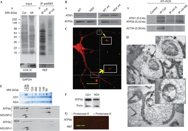
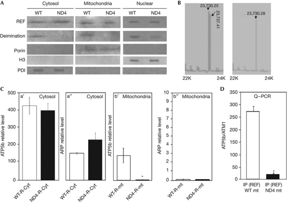
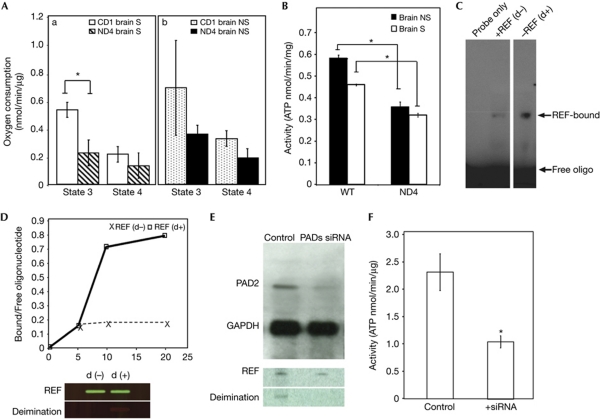
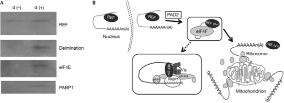
Deimination refers to conversion of protein-bound arginine into citrulline. An mRNA carrier, RNA binding export factor (REF), present on mitochondria undergoes loss of deimination with impaired ATP5b mRNA transport in ND4 mice (model of multiple sclerosis) compared with the controls. We present evidence of (1) reduced ATP5b mRNA binding strength of non-deiminated REF compared with deiminated REF, (2) impaired ATP5b mRNA transport in ND4 mice and (3) reduced mitochondrial ATP synthase activity on inhibition of deimination in PC12 cells. Impaired deimination of REF and defect in mitochondrial mRNA transport are critical factors in mitochondrial dysfunction in ND4 mice.


Conflict of interest statement

The authors declare that they have no conflict of interest.

Figures

Figure 1
Figure 1
REF and ATP5b status on mitochondrial fraction. (A) Demonstration of RNA binding export factor (REF) in mitochondrial fraction. AntiREF immunoprecipitation (IP) from cytosolic or mitochondrial fraction. (a) The input retinal cytosolic and brain mitochondrial protein extracts were separated on a 10% SDS–PAGE and detected with antiCOX IV and glyceraldehyde-3-phosphate dehydrogenase (GAPDH) as indicated. (b) IP products from inputs as shown in panel a were gel fractionated and visualized using silver staining. Bottom panel shows recovery of REF using western analysis. (B) Detection of ATP5b and ATM1 mRNA by reverse transcription–polymerase chain reaction (RT–PCR). (a) ATM1 and ATP5b with wild-type (WT) and ND4 mitochondrial (mt) fraction. (b) Detection of ATP5b, ATM and actin in normal mouse complementary DNA derived from three IP products: Retinal cytosolic (cyt), non-synaptic (mt-NS) and synaptic (mt-S) mitochondria as indicated were carried out using appropriate primer pairs. Control represents RT–PCR from an IP performed with an antibody unrelated to any known mammalian protein. Bottom panel shows amplification of actin used as a control. The product size has been provided in the parentheses. (C) Localization of REF on the mitochondrial surface. Primary hippocampal neuron culture was probed with Porin (red) and REF (green) antibodies. Merged figure has been shown. White boxes indicated co-localization of REF and Porin. Z axis and magnified view indicated by yellow arrows. Scale bar, 20 μm. (D) Electron microscopy in detection of REF at mitochondrial surface. Upper and lower panel indicate CD1 and ND4 mice sections, respectively. Scale bar, 0.2 μm. Arrow and arrowhead indicate the region shown in the inset and a mitochondrial gold particle, respectively. (E) Blue native gel electrophoresis of mitochondrial protein extract from CD1 and ND4 mice as indicated (upper panel). Bottom panel shows western blot analyses from a posttransfer two-dimensional SDS–PAGE. Arrowhead indicate NADH dehydrogenase ubiquinone iron-sulphur protein 4 (NDUSF4); proteins were probed with antibodies as indicated. (F) Western blot analyses of mt proteins from CD1 and ND4 mice with ATP5b and Porin as indicated. (G) Control and mt subjected to digestion with Proteinase K as indicated probed for REF and ATP5b, with secondary antibodies coupled with IR-700, IR-800 and scanned on an Odyssey infrared imaging system (Li-Cor Biosciences, Lincoln, NE). MW, molecular weight.
Figure 2
Figure 2
Comparison of ATP5b expression and ATP synthase activity between wild-type and ND4 mice. (A) Loss of RNA binding export factor (REF) deimination in ND4 mice. Western analysis of deimination and REF in the cytosol, nuclear fraction and mitochondria of wild-type (WT) and ND4 mice as indicated. The deimination status of REF was detected using anticitrulline antibody (bottom panel), blots were probed with Porin, histone H3 and protein disulphide isomerase (PDI) as indicated. (B) Analysis of intact purified REF using mass spectrometry on a matrix-assisted laser desorption/ionization–time of flight device in linear mode. Arrowheads indicated non-deiminated form, arrows indicated deiminated form with m/z ratios. Cyt, retinal cytosolic soluble proteins; mt, mitochondria; IP, immunoprecipitation. (C) ATP5b transcript levels were quantified and normalized by ATM1 mRNA levels by real-time reverse transcription–polymerase chain reaction (PCR) and compared between control (WT) and ND4 mice as indicated. (a′, a″) ATP5b and actin-related protein (ARP) mRNA levels in retinal cytosolic fraction and (b′, b″) mitochondrial fraction as indicated. ARP mitochondrial level is ∼2,000-fold less compared with ATP5b or cytosolic ARP levels. (D) Comparison of REF and ATP5b association between WT (CD1) and ND4 mice. ATP5b expression from complementary DNA recovered by reverse transcription from IP products: wild-type mitochondria (WT mt) and ND4 mitochondria (ND4 mt) as indicated was quantified by real-time PCR and normalized by ATM1 levels. NS, non-synaptic mitochondria; S, synaptic mitochondria. All comparisons were made from at least three independent measurements with analysis of variance expressed as mean±s.d. All data were subjected to two-tailed paired t-test against controls (and were considered significant *P⩽0.05).
Figure 3
Figure 3
Deiminated REF in regulation of ATP synthase expression. (A) Comparison of mean mitochondrial states 3 and 4 respiration rates between wild-type (WT; CD1) and ND4 mice. (a) Respiration rates (states 3 and 4 as indicated) in synaptic (S) mitochondria isolated from ND4 (n=4) and CD1 (n=6) brains. (b) Respiration rates (states 3 and 4 as indicated) in non-synaptic (NS) mitochondria isolated from ND4 (n=4) and CD1 (n=6) brains. Rate of oxygen consumption was measured in the presence and in the absence of ADP. (B) Comparison of mean mitochondrial ATP synthase activity between WT (CD1) and ND4 mice. Isolated mitochondria from brain non-synaptic (NS, solid column) and synaptosomes (S, hollow column) were applied to ATP synthase measurement as indicated. (C) Electrophoretic mobility shift assay (EMSA) analysis of deiminated REF using an oligonucleotide probe 5′-TGGGCAGAATCATGAATGTC-3′. EMSA of non-deiminated (d−) and deiminated (d+) REF as indicated in lane 2 and 3; the control lane was loaded only with probe. (D) Time course was generated by the ratio of bound and free probe (bound/free oligonucleotide) and incubation time and compared between non-deiminated REF (d−; cross and dashed line) and deiminated REF (d+; square; solid line). Protein loading from an EMSA gel was probed for REF and its deimination status (lower panel) using antiREF and anticitrulline with secondary antimouse and antirabbit coupled with IR-700 and IR-800 scanned on an Odyssey infrared imaging system (Li-Cor Biosciences, Lincoln, NE). (E) The mRNA expression after siRNA inhibition. Representative PAD2 and GAPDH protein as indicated were compared between the control and transfected cells. The expression level was quantified by ImageJ program (supplementary Fig S4D online). Control and PAD siRNA inhibited product were probed with antiREF and anticitrulline antibody, respectively (bottom panel). (F) The ATP synthase activity of PC12 cell line after PAD2 and PAD4 siRNA treatment. Control indicates the cells transfected with nonspecific negative siRNA, + siRNAs indicates the cells transfected with PAD2 and PAD4 siRNAs. All comparisons were made from at least three independent measurements with analysis of variance expressed as mean±s.d. All data were subjected to two-tailed paired or unpaired t-test with respect to controls (*), *P⩽0.05. ATP, adenosine triphosphate; GAPDH, glyceraldehyde-3-phosphate dehydrogenase; REF, RNA binding export factor; siRNA, short interfering RNA.
Figure 4
Figure 4
Association of deiminated REF with protein translational machinery components. (A) Western analyses of antiREF (Refbp2) immunoprecipitation products with deiminated REF (d+) and control non-deiminated REF (d−) as bait. (B) Schematic diagram of REF mediate mRNA export at mitochondrial surface. The REF (indicated) exports polyadenylated cargo from the nucleus. REF becomes additionally deiminated (indicated as d+) by PAD2 and recruits members of protein translation initiation complex elf4F, facilitate the mRNA (ATP5b) transport to mitochondrial surface; details of REF interacting protein complex have been shown in the enlarged box. REF, RNA binding export factor.

Similar articles

Cited by

References

    1. Franke J, Reimann B, Hartmann E, Kohlerl M, Wiedmann B (2001) Evidence for a nuclear passage of nascent polypeptide-associated complex subunits in yeast. J Cell Sci 114: 2641–2648 - PubMed
    1. Ding D, Dave KR, Bhattacharya SK (2009) On message ribonucleic acids targeting to mitochondria. Biochem Insights 2: 1–11
    1. Fujiki M, Verner K (1993) Coupling of cytosolic protein synthesis and mitochondrial protein import in yeast. Evidence for cotranslational import in vivo. J Biol Chem 268: 1914–1920 - PubMed
    1. Sylvestre J, Margeot A, Jacq C, Dujardin G, Corral-Debrinski M (2003a) The role of the 3′ untranslated region in mRNA sorting to the vicinity of mitochondria is conserved from yeast to human cells. Mol Biol Cell 14: 3848–3856 - PMC - PubMed
    1. Dutta R et al. (2006) Mitochondrial dysfunction as a cause of axonal degeneration in multiple sclerosis patients. Ann Neurol 59: 478–489 - PubMed

Publication types

MeSH terms