. 2012 Jun;11(6):M111.014233.
doi: 10.1074/mcp.M111.014233. Epub 2012 Jan 19.

Interaction proteomics identify NEURL4 and the HECT E3 ligase HERC2 as novel modulators of centrosome architecture

Affiliations

Interaction proteomics identify NEURL4 and the HECT E3 ligase HERC2 as novel modulators of centrosome architecture

Abdallah K Al-Hakim et al. Mol Cell Proteomics. 2012 Jun.

Abstract

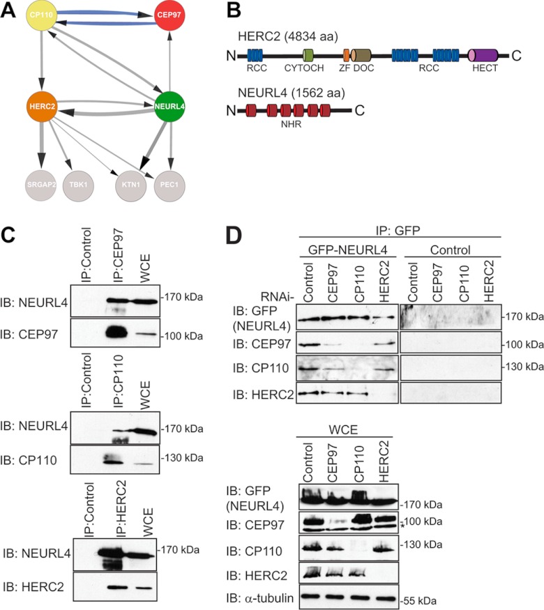
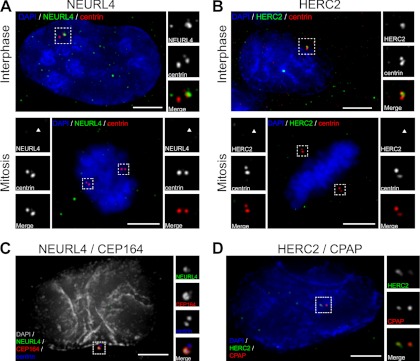
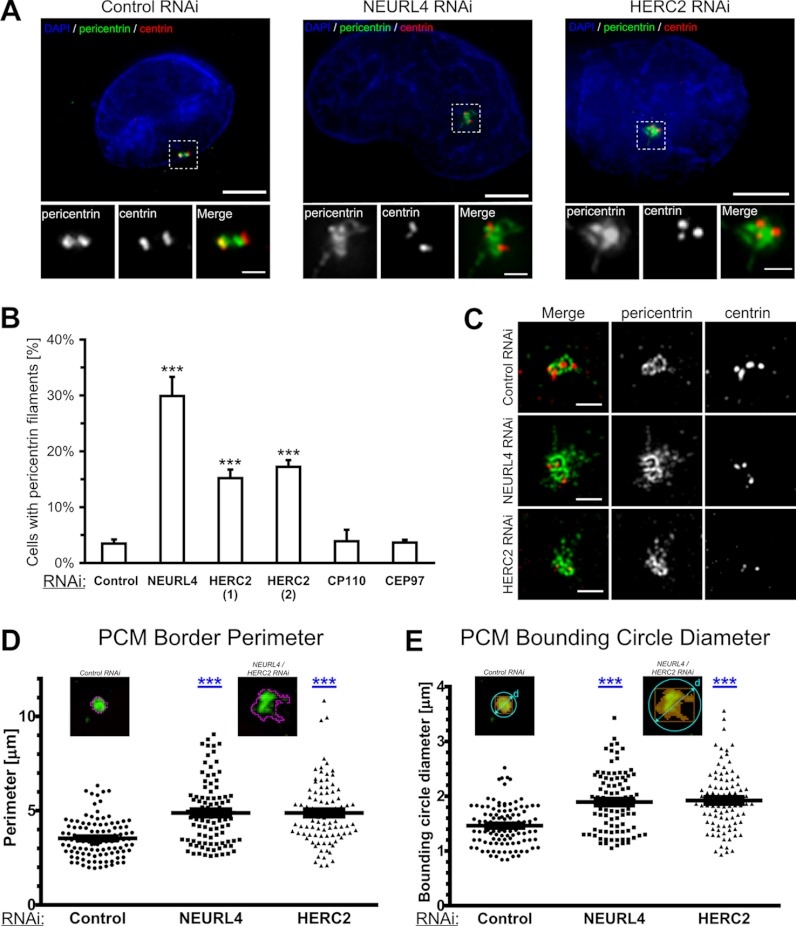
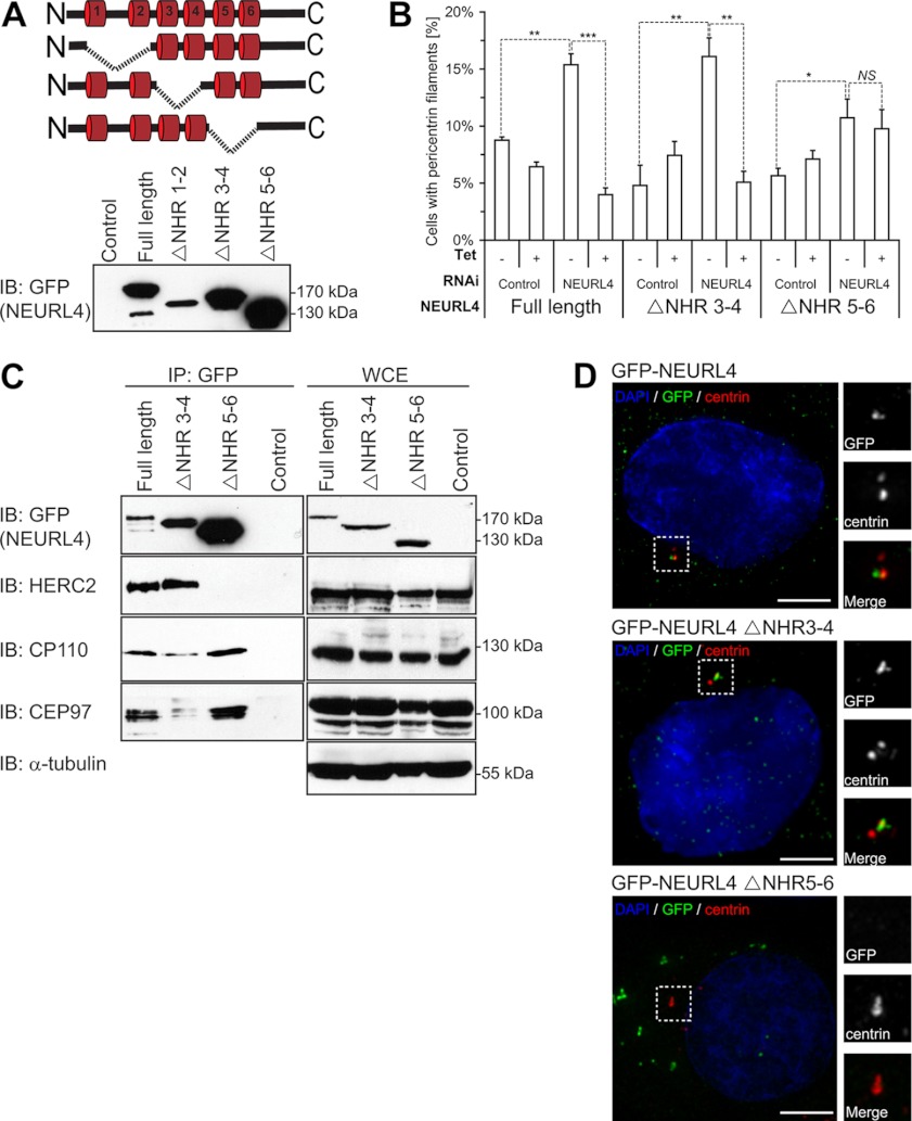
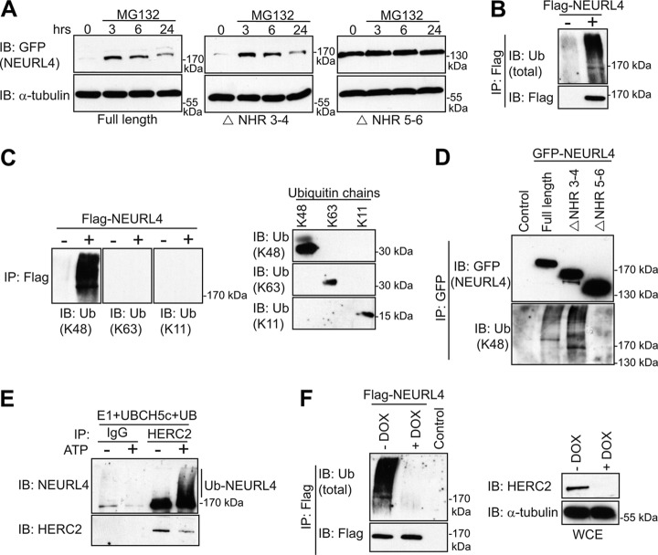
Centrosomes are composed of a centriole pair surrounded by an intricate proteinaceous matrix referred to as pericentriolar material. Although the mechanisms underpinning the control of centriole duplication are now well understood, we know relatively little about the control of centrosome size and shape. Here we used interaction proteomics to identify the E3 ligase HERC2 and the neuralized homologue NEURL4 as novel interaction partners of the centrosomal protein CP110. Using high resolution imaging, we find that HERC2 and NEURL4 localize to the centrosome and that interfering with their function alters centrosome morphology through the appearance of aberrant filamentous structures that stain for a subset of pericentriolar material proteins including pericentrin and CEP135. Using an RNA interference-resistant transgene approach in combination with structure-function analyses, we show that the association between CP110 and HERC2 depends on nonoverlapping regions of NEURL4. Whereas CP110 binding to NEURL4 is dispensable for the regulation of pericentriolar material architecture, its association with HERC2 is required to maintain normal centrosome integrity. NEURL4 is a substrate of HERC2, and together these results indicate that the NEURL4-HERC2 complex participates in the ubiquitin-dependent regulation of centrosome architecture.


Figures

Fig. 1.
Fig. 1.
Identification of novel CP110 interacting proteins. A, the interaction network for NEURL4, HERC2, CP110, and CEP97 was prepared using Cytoscape software (–58). The parameters used for the indicated interactions were: minimum number of unique peptides = 2; maximum frequency across the database = 20; minimum Mascot score = 60; ≥40 total peptide numbers; remove all hits detected in any one of control samples; included proteins detected in at least two-thirds of the biological replicates for each bait. The control samples were as follows: three of the control runs included data from control IP in HEK293 cells where rabbit IgG coupled to protein G-Sepharose resin was used. The other remaining control runs included data from IPs in HEK293 Flp-In T-REx cell lines expressing empty vector with FLAG-M2-agarose resin. The thicknesses of the connecting lines are proportional to the average number of total peptides associated with the interaction. The gray connecting lines indicate new interactions, whereas the blue connecting lines indicate previously reported interactions. B, domain structure of HERC2 and NEURL4. RCC, regulator of chromosome condensation (amino acids 514–727, 2959–3327, and 3952–4319); CYTOCH, cytochrome b5 heme-binding domain (amino acids 1207–1283); ZF, zinc finger (amino acids 2702–2749); DOC, Anaphase-promoting complex, subunit 10 (amino acids 2759–2930); HECT, homologous to the E6-AP C terminus (amino acids 4457–2794); NHR, neuralized homology region (amino acids 41–207, 317–484, 520–686, 716–884, 913–1086, and 1131–1294). C, endogenously expressed CEP97, CP110, and HERC2 were immunoprecipitated from HEK293T WCE with their own respective antibodies. As a control, IP was performed in parallel with a nonspecific rabbit IgG. Immunoprecipitated proteins were subjected to immunoblot analysis using the CEP97, CP110, HERC2, and NEURL4 antibodies. D, U2OS cells stably expressing GFP-NEURL4 were transfected with esiRNA targeting CEP97, CP110, HERC2, or luciferase (negative control). 72 h post-transfection, cells were harvested, and GFP-NEURL4 was immunoprecipitated using anti-GFP antibody (top panel). Right top panel, a GFP control IP was performed in U2OS cells expressing an empty vector and immunoblotted with GFP, CEP97, CP110, and HERC2 antibodies. The immune complexes were subjected to immunoblot analysis with CEP97, CP110, HERC2, and GFP antibodies. WCE were blotted as a loading control (bottom panel). The protein size (kDa) is indicated for each immunoblot. The nonspecific cross-reactive bands are indicated by an asterisk. C and D, HERC2 is a 528-kDa protein and runs as a very high molecular mass band, and there is no molecular mass standard in that region of the gel. IB, immunoblot.
Fig. 2.
Fig. 2.
NEURL4 and HERC2 are novel centrosomal proteins. A and B, U2OS cells were labeled for DNA (blue; DAPI), centrin (red), and NEURL4 (green) (A) or DNA (blue), centrin (red), and HERC2 (green) (B). Interphase cells are shown in the top panels. The images show co-localization of NEURL4 (A) and HERC2 (B) with centriolar marker centrin (scale bar, 5 μm). Both NEURL4 and HERC2 are absent from the centrosomes in mitosis (bottom panels). The insets are 2-fold magnifications of the outlined areas to better visualize the centrosomal regions. C, U2OS cells were sequentially labeled for NEURL4 (green) and CEP164 (red), followed by labeling for centrin (blue) and DNA (gray, DAPI). NEURL4 co-localizes with Cep164 to the distal appendages of mature centriole. D, U2OS cells were labeled for DNA (blue; DAPI), CPAP (red), and HERC2 (green). HERC2 co-localization with CPAP, a marker of the proximal region of centrioles (scale bar, 5 μm). The insets are 2-fold magnifications of the outlined areas to better visualize the centrosomal regions. DAPI, 4′,6′-diamino-2-phenylindole.
Fig. 3.
Fig. 3.
NEURL4 and HERC2 regulate centrosome morphology. A, U2OS cells were transfected with siGENOME nontargeting siRNA #1, NEURL4 siRNA, or HERC2 esiRNA-2 and labeled for DNA (blue; DAPI), centrin (red), and pericentrin (green). The cells were imaged using conventional deconvolution microscopy. NEURL4 depletion caused abnormalities in pericentriolar material, shown in more detail in the insets. The insets are magnifications of the outlined centrosomal regions. Scale bars, 5 μm. B, U2OS cells were transfected with luciferase (control), HERC2(1), HERC2(2), CP110, CEP97 esiRNA, or NEURL4 siRNA. The presence of pericentrin filaments was quantified 72 h post-transfection. The graph shows the averages ± S.E. of the number of cells with pericentrin filaments, obtained in three independent experiments. At least 100 cells were analyzed under each condition. The increase in the number of pericentrin filaments is statistically significant. ***, p value < 0.0001. C, cells treated as described in A were imaged on a structured-illumination microscope. High resolution images of the centrosome regions reveal fine details of the filamentous structures, induced by NEURL4 or HERC2 depletion. Scale bars, 1 μm. D and E, wild type and filamentous centrosomes were segmented, based on an adaptive threshold. The PCM border perimeter (D) and the PCM bounding circle diameter (E) of each detected object (see insets) were then analyzed. The increase in both parameters was highly significant. ***, p value < 0.0001. Cut-off for considering a centrosome as filamentous was set at 5 μm for border perimeter and 2 μm for bounding circle diameter. At least 30 centrosomes were measured in three independent experiments for each condition. DAPI, 4′,6′-diamino-2-phenylindole.
Fig. 4.
Fig. 4.
Structure-function analysis of NEURL4. A, schematic representation of the different deletion constructs generated for GFP-NEURL4 protein (top panel). Stable U2OS cell lines were generated that expressed each of these deletion constructs, and protein expression was confirmed by immunoblot analysis with the GFP antibody (bottom panel). B, U2OS cell expressing inducible GFP-tagged NEURL4 or NHR deletion mutants were transfected with luciferase esiRNA or esiRNA targeting the 3′-UTR region of NEURL4. Presence of pericentrin filaments was quantified 48 h after induction with 1 μg/ml tetracycline (where indicated) and 72 h after RNAi transfection. The graph shows the averages ± S.E. of the number of cells with pericentrin filaments, obtained in three independent experiments. At least 100 cells were analyzed under each condition. t test results comparing the indicated data sets are shown in the graph. *, p value < 0.05; **, p value < 0.001; ***, p value < 0.0001. NS, nonsignificant difference. C, U2OS stable cell lines expressing the GFP-tagged NEURL4, NEURL4 ΔNHR3–4, and NEURL4 ΔNHR5–6 constructs were induced for expression with 1 μg/ml tetracycline for 16–18 h before harvesting. The exogenously expressed GFP-NEURL4 was immunoprecipitated using GFP antibodies, and the immune complexes were subjected to immunoblot analysis with antibodies against GFP, HERC2, CP110, and CEP97 (left panel). A control IP was performed in parallel in U2OS stable cell lines expressing an empty vector. WCEs were immunoblotted with antibodies to the same proteins as well as the α-tubulin as a loading control (right panel). The control samples were WCEs from U2OS cell line expressing empty vector. HERC2 is a 528-kDa protein and runs as a very high molecular mass band, and there is no molecular mass standard in that region of the gel. D, U2OS cells expressing inducible GFP-tagged NEURL4, NEURL4 ΔNHR3–4, and NEURL4 ΔNHR5–6 were treated with 1 μg/ml tetracycline for 48 h to induce expression. The cells were labeled for DNA (blue; DAPI), pericentrin (red), and GFP (green). Scale bars, 5 μm. GFP-NEURL4 ΔNHR3–4 is co-localized with pericentrin, whereas GFP-NEURL4 ΔNHR5–6 is excluded from the centrosomal region. The insets are magnifications of the outlined regions. The protein size (kDa) is indicated for each immunoblot. DAPI, 4′,6′-diamino-2-phenylindole; IB, immunoblot.
Fig. 5.
Fig. 5.
NEURL4 is ubiquitylated in a HERC2-dependent manner. A, expression of the GFP-tagged NEURL4, NEURL4 ΔNHR3–4, and NEURL4 ΔNHR5–6 was induced with tetracycline in the U2OS stable cells lines. 24 h post-induction, the cells were treated with 10 μm MG132. The cells were lysed at 3, 6, and 24 h post-MG132 additions. The protein levels of NEURL4 were determined by immunoblotting with the GFP antibody, and α-tubulin immunoblot was performed as a loading control. B, FLAG-NEURL4 protein was immunoprecipitated from HEK293T cells and immunoblotted with total ubiquitin antibody. As a control, we performed the experiment in parallel with cells transfected with an empty vector (right panel). C, left panel, as in B but immunoblotted with antibodies specific to K11, K48, and K63 ubiquitin chains. Right panel, 250 ng each of tetra-ubiquitin K48, tetra-ubiquitin K63, or di-ubiquitin K11 were subjected to SDS-PAGE, transferred to nitrocellulose membranes, and immunoblotted with ubiquitin antibodies specific for K48, K11, or K63 ubiquitin chains. D, WCEs from U2OS cell lines stably expressing different GFP-NEURL4 constructs were subjected to immunoprecipitation using the GFP antibody. Immunoprecipitated proteins were analyzed by SDS-PAGE and immunoblotted with GFP antibody or ubiquitin antibody specific to K48 polyubiquitin chains. E, endogenously expressed HERC2 was immunopurified from U2OS cells using HERC2 antibody conjugated to protein G-Sepharose. Although the protein was still on the resin, UBE1, UBCH5c, and ATP were added to initiate the ubiquitylation assay. The assay was terminated by the addition of SDS buffer, and the eluted proteins were subjected to immunoblot analysis with antibodies against NEURL4 and HERC2. As a control, IP was performed in parallel with a nonspecific rabbit IgG. F, U2OS cells stably expressing doxycycline-inducible shHERC2 were transfected with FLAG-NEURL4. 96 h post-shHERC2 induction, the cells were lysed, subjected to FLAG-IP, and immunoblotted with antibodies against FLAG and total ubiquitin. As a control, the IP was performed in parallel with cells transfected with an empty vector. The efficiency of knockdown and loading controls are shown in the lower panel. The protein size (kDa) is indicated for each immunoblot. E and F, HERC2 is a 528-kDa protein and runs as a very high molecular mass band, and there is no molecular mass standard in that region of the gel. IB, immunoblot; Ub, ubiquitin; DOX, doxycycline.

Similar articles

Cited by

References

    1. Andersen J. S., Wilkinson C. J., Mayor T., Mortensen P., Nigg E. A., Mann M. (2003) Proteomic characterization of the human centrosome by protein correlation profiling. Nature 426, 570–574 - PubMed
    1. Nigg E. A., Raff J. W. (2009) Centrioles, centrosomes, and cilia in health and disease. Cell 139, 663–678 - PubMed
    1. Palazzo R. E., Vogel J. M., Schnackenberg B. J., Hull D. R., Wu X. (2000) Centrosome maturation. Curr. Top. Dev. Biol. 49, 449–470 - PubMed
    1. Conduit P. T., Brunk K., Dobbelaere J., Dix C. I., Lucas E. P., Raff J. W. (2010) Centrioles regulate centrosome size by controlling the rate of Cnn incorporation into the PCM. Curr. Biol. 20, 2178–2186 - PubMed
    1. Decker M., Jaensch S., Pozniakovsky A., Zinke A., O'Connell K. F., Zachariae W., Myers E., Hyman A. A. (2011) Limiting amounts of centrosome material set centrosome size in C. elegans embryos. Curr. Biol. 21, 1259–1267 - PubMed

Publication types

MeSH terms

LinkOut - more resources