

In vivo comparison of arterial lumen dimensions assessed by co-registered three-dimensional (3D) quantitative coronary angiography, intravascular ultrasound and optical coherence tomography
- PMID: 22261998
- PMCID: PMC3463784
- DOI: 10.1007/s10554-012-0016-6
In vivo comparison of arterial lumen dimensions assessed by co-registered three-dimensional (3D) quantitative coronary angiography, intravascular ultrasound and optical coherence tomography
Abstract
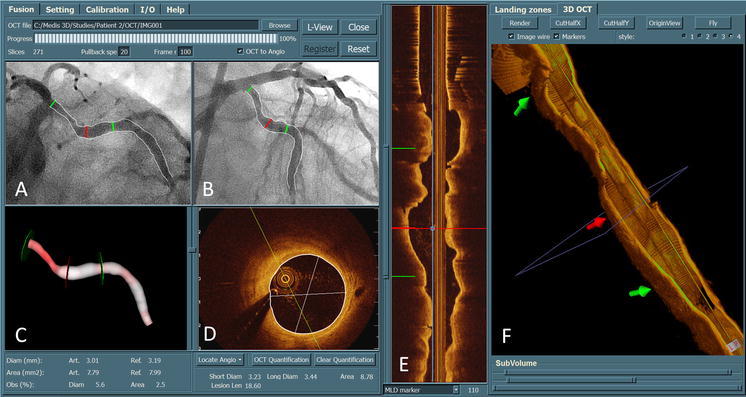
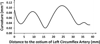
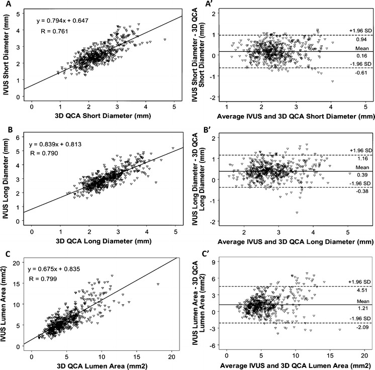
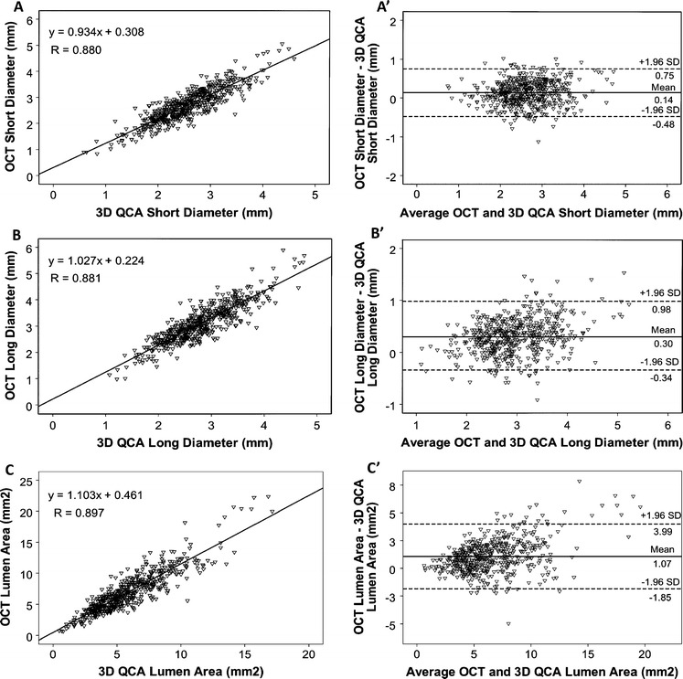
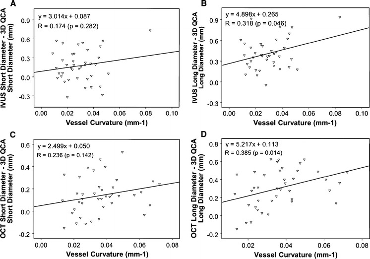
This study sought to compare lumen dimensions as assessed by 3D quantitative coronary angiography (QCA) and by intravascular ultrasound (IVUS) or optical coherence tomography (OCT), and to assess the association of the discrepancy with vessel curvature. Coronary lumen dimensions often show discrepancies when assessed by X-ray angiography and by IVUS or OCT. One source of error concerns a possible mismatch in the selection of corresponding regions for the comparison. Therefore, we developed a novel, real-time co-registration approach to guarantee the point-to-point correspondence between the X-ray, IVUS and OCT images. A total of 74 patients with indication for cardiac catheterization were retrospectively included. Lumen morphometry was performed by 3D QCA and IVUS or OCT. For quantitative analysis, a novel, dedicated approach for co-registration and lumen detection was employed allowing for assessment of lumen size at multiple positions along the vessel. Vessel curvature was automatically calculated from the 3D arterial vessel centerline. Comparison of 3D QCA and IVUS was performed in 519 distinct positions in 40 vessels. Correlations were r = 0.761, r = 0.790, and r = 0.799 for short diameter (SD), long diameter (LD), and area, respectively. Lumen sizes were larger by IVUS (P < 0.001): SD, 2.51 ± 0.58 mm versus 2.34 ± 0.56 mm; LD, 3.02 ± 0.62 mm versus 2.63 ± 0.58 mm; Area, 6.29 ± 2.77 mm(2) versus 5.08 ± 2.34 mm(2). Comparison of 3D QCA and OCT was performed in 541 distinct positions in 40 vessels. Correlations were r = 0.880, r = 0.881, and r = 0.897 for SD, LD, and area, respectively. Lumen sizes were larger by OCT (P < 0.001): SD, 2.70 ± 0.65 mm versus 2.57 ± 0.61 mm; LD, 3.11 ± 0.72 mm versus 2.80 ± 0.62 mm; Area 7.01 ± 3.28 mm(2) versus 5.93 ± 2.66 mm(2). The vessel-based discrepancy between 3D QCA and IVUS or OCT long diameters increased with increasing vessel curvature. In conclusion, our comparison of co-registered 3D QCA and invasive imaging data suggests a bias towards larger lumen dimensions by IVUS and by OCT, which was more pronounced in larger and tortuous vessels.
Figures
References
-
- Tu S, Jing J, Holm NR, Onsea K, Zhang T, Adriaenssens T, Dubois C, Desmet W, Thuesen L, Chen Y, Reiber JHC (2011) In vivo Assessment of bifurcation optimal viewing angles and bifurcation angles by three-dimensional (3D) quantitative coronary angiography. Int J Cardiovasc imaging. Epub Ahead of Print. doi: 10.1007/s10554-011-9996-x - PMC - PubMed
Publication types
MeSH terms
LinkOut - more resources
Full Text Sources
Other Literature Sources
Medical
Research Materials
