. 2012 Apr 15;302(8):C1174-88.
doi: 10.1152/ajpcell.00450.2011. Epub 2012 Jan 18.

Expression of Na+-D-glucose cotransporter SGLT2 in rodents is kidney-specific and exhibits sex and species differences

Affiliations

Expression of Na+-D-glucose cotransporter SGLT2 in rodents is kidney-specific and exhibits sex and species differences

Ivan Sabolic et al. Am J Physiol Cell Physiol. .

Abstract

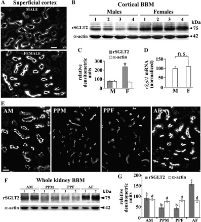
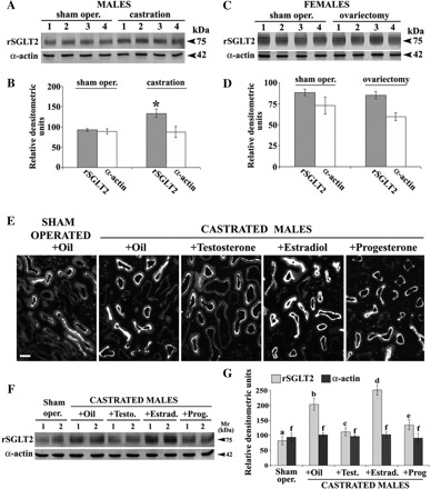
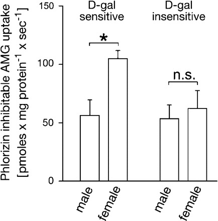
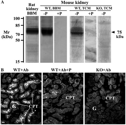
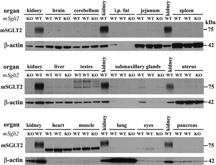
With a novel antibody against the rat Na(+)-D-glucose cotransporter SGLT2 (rSGLT2-Ab), which does not cross-react with rSGLT1 or rSGLT3, the ∼75-kDa rSGLT2 protein was localized to the brush-border membrane (BBM) of the renal proximal tubule S1 and S2 segments (S1 > S2) with female-dominant expression in adult rats, whereas rSglt2 mRNA expression was similar in both sexes. Castration of adult males increased the abundance of rSGLT2 protein; this increase was further enhanced by estradiol and prevented by testosterone treatment. In the renal BBM vesicles, the rSGLT1-independent uptake of [(14)C]-α-methyl-D-glucopyranoside was similar in females and males, suggesting functional contribution of another Na(+)-D-glucose cotransporter to glucose reabsorption. Since immunoreactivity of rSGLT2-Ab could not be detected with certainty in rat extrarenal organs, the SGLT2 protein was immunocharacterized with the same antibody in wild-type (WT) mice, with SGLT2-deficient (Sglt2 knockout) mice as negative control. In WT mice, renal localization of mSGLT2 protein was similar to that in rats, whereas in extrarenal organs neither mSGLT2 protein nor mSglt2 mRNA expression was detected. At variance to the findings in rats, the abundance of mSGLT2 protein in the mouse kidneys was male dominant, whereas the expression of mSglt2 mRNA was female dominant. Our results indicate that in rodents the expression of SGLT2 is kidney-specific and point to distinct sex and species differences in SGLT2 protein expression that cannot be explained by differences in mRNA.


Figures

Fig. 1.
Fig. 1.
Immunoreactivity of antibody against rat SGLT2 (rSGLT2-Ab) in kidneys and small intestine of female rats. A: optimal conditions for Western blots with total cell membranes (TCM) from rat kidney cortex. For SDS-PAGE, isolated TCM were prepared in Laemmli buffer with (+β-ME) or without (−β-ME) β-mercaptoethanol and heated for 5 min at 95°C, 15 min at 65°C, or 30 min at 37°C. In all conditions, only a broad ∼75-kDa protein band was labeled. B: Western blot of TCM isolated from the rat kidney cortex (K) or mucosa of the small intestine (jejunum; I) with rSGLT2-Ab (Ab) or with the antigenic peptide-blocked rSGLT2-Ab (Ab+P). Each lane in A and B contained 60 μg of protein. C: Western blot of brush-border membrane (BBM) isolated from whole rat kidneys with the native (Ab) or peptide-blocked (Ab+P) rSGLT2-Ab. Each lane contained 40 μg of protein. Denaturation of the membranes for B and C, as well as in all further experiments, was performed for 15 min at 65°C (−β-ME). D: immunostaining of kidney cortex (K) and small intestine (I) with rSGLT2-Ab or with the peptide-blocked rSGLT2-Ab (K+P). In kidney cortex (K), rSGLT2-Ab strongly stained the brush border of proximal tubules, while the brush border of villi in intestine remained unstained. After preabsorption of rSGLT2-Ab with the antigenic peptide, no staining of renal cortical tubules was observed (K+P). G, glomerulus; PT, proximal tubule; S1, S1 segment; S2, S2 segment. E: zonal differences: rSGLT2-Ab immunoreactivity in superficial and deep cortex and in outer stripe. rSGLT2-Ab exclusively stained the BBM of cortical proximal tubules; staining in the S1 segments (asterisks) was stronger compared with the S2 segments (arrows). Data represent findings in 3 female rats. G, glomerulus. S3 segments in the outer stripe (arrowheads) were not stained. Bars in D and E, 20 μm. F: Western blot of TCM isolated from the rat kidney cortex (CTX) or outer stripe (OS). The experiment was performed as in B. Prominent staining of the 75-kDa protein band was only observed in cortical membranes.
Fig. 2.
Fig. 2.
Immunolocalization of rSGLT2 and cell adhesion molecule CAM105 in proximal tubular segments. Staining with rSGLT2-Ab (A, C) and CAM105-Ab (B, D) was performed in consecutive sections of rat kidney cortex (A, B) and outer stripe (C, D). In cortex (A), rSGLT2-Ab stained the high brush border of S1 segments and the low brush border of S2 segments. In accordance with previous data (37), these segments can be distinguished by BBM staining with CAM105-Ab, which is strong in the S1 segment (B, arrowheads) and weak or absent in the S2 segment (B). In the outer stripe, the brush border of S3 segments was negative for rSGLT2 (C) and strongly positive for CAM105 (D, arrowheads). Data represent findings in 2 female rats. Cell membranes of the glomerular (G) and peritubular capillaries (arrows, B and D) were also positive for CAM105. Bar, 20 μm.
Fig. 3.
Fig. 3.
Sex-dependent expression of rSGLT2 in kidneys of adult (A–D) and prepubertal (E–G) rats. A: immunostaining with rSGLT2-Ab in cortical proximal tubules of an adult male (M) and female (F) rat. The staining intensity of the proximal tubule brush border in the M was weaker than in the F. Data represent findings in 2 animals of each sex. Bar, 20 μm. B: abundance of rSGLT2 protein and α-actin in isolated renal cortical BBM from 4 adult M and F. Each lane contained 40 μg protein. C: densitometric evaluation of the bands shown in B; the rSGLT2-related 75-kDa protein band was ∼3-fold stronger in adult F compared with adult M (*P < 0.05), whereas the α-actin-related 42-kDa protein band showed no sex differences. D: expression of rSglt2 mRNA in kidneys of adult M and F as estimated by real-time RT-PCR (N = 5 for each sex). No significant sex difference was detected (n.s., not significant). E: comparison of rSGLT2-Ab immunostaining in the renal cortex of adult M (AM), prepubertal M (PPM) and F (PPF), and adult F (AF) rats. Whereas adult animals exhibited stronger staining in females, the staining in prepubertal rats was much weaker than in the adult rats and sex independent. In control experiments, the staining in both adult and prepubertal rats was abolished when rSGLT2-Ab was preincubated with the antigenic peptide (not shown). Data represent findings in 3 rats of each sex. Bar, 20 μm. F: representative immunoblots of rSGLT2 and α-actin in BBM isolated from the whole kidneys of adult (AM and AF) and prepubertal (PPM and PPF) rats. Immunoblots were performed with 60 μg protein/lane. G: densitometric quantification of the rSGLT2-related 75-kDa protein band (N = 4 for each group). In PPM and PPF the rSGLT2-related bands showed similar intensity; they were ∼50% weaker than in adult M and ∼70% weaker than in adult F. The α-actin band was age- and sex independent. Statistics (ANOVA/Duncan test): P < 0.05: a/b, a/c, and b/c; n.s.: b/b and d/d.
Fig. 4.
Fig. 4.
Effects of gonadectomy in male (M) and female (F) rats (A–D) and of sex hormone treatment in castrated M (E–G) on renal expression of rSGLT2 protein. A: Western blots of rSGLT2 and α-actin in isolated renal cortical BBM from 4 sham-operated and 4 castrated M. B: densitometric evaluation of the bands shown in A; the rSGLT2-related 75-kDa protein band increased ∼50% after castration (vs. sham-operated control rats, *P < 0.05), whereas the α-actin band was unaffected by castration. C: Western blots of rSGLT2 and α-actin in isolated renal cortical BBM from 4 sham-operated and 4 ovariectomized F. D: densitometric quantification of the bands shown in C; both the rSGLT2- and α-actin-related bands remained unchanged after ovariectomy. Blots were performed with 40 μg protein/lane. E: immunostaining with rSGLT2-Ab in cryosection of the kidney cortex from oil-treated sham-operated adult M and castrated males treated with oil, testosterone, estradiol, or progesterone. In all cases, the staining was abolished after block of the antibody with the immunizing peptide (not shown). Data represent findings in 3 rats from each experimental group. Bar, 20 μm. F: representative Western blot of rSGLT2 in TCM from the kidney cortex from sham-operated, castrated, and hormone-treated castrated M. Blots were performed with 60 μg protein/lane. G: densitometric quantification of the bands shown in F and collected from 2 similar experiments with independent membrane preparations (N = 4 in each group). Statistics for the 75-kDa band (ANOVA/ Duncan test): P < 0.05: a/b, a/d, a/e, b/c, b/d, and b/e; n.s.: a/c, c/e, and f/f. The density pattern of the 75-kDa protein band in castrated M treated with oil or sex hormones resembled the immunostaining data, i.e., the band was upregulated by castration (2.5-fold), whereas in castrated M it was strongly diminished by testosterone (∼50%), increased by estradiol (∼20%), and decreased by progesterone (∼33%) treatment. The 42-kDa α-actin band was not affected by castration or hormone treatment.
Fig. 5.
Fig. 5.
d-Galactose (d-gal)-sensitive and d-gal-insensitive α-methyl-d-glucopyranoside (AMG) uptake into BBM vesicles isolated from the whole kidneys of male and female rats. The inward Na+ gradient-driven uptake of 0.1 mM [14C]AMG was measured by rapid filtration technique in the absence of inhibitors, in the presence of 20 mM d-gal, and in the absence or presence of 0.2 mM phlorizin. AMG uptake in the presence of sodium that can be inhibited by phlorizin represents total Na+-d-glucose cotransport. The d-gal-inhibitable fraction is supposed to represent high-affinity Na+-d-glucose cotransport mediated by rSGLT1 (d-gal sensitive), whereas the d-gal insensitive, phlorizin-inhibitable AMG uptake (d-gal insensitive) represents the low-affinity Na+-d-glucose cotransport, which may be mediated by rSGLT2 and NaGLT1. BBM vesicles from the female cortex exhibited ∼85% higher d-gal-inhibitable AMG uptake compared with that in males, whereas the remaining, phlorizin-sensitive, d-gal-insensitive AMG uptake in vesicles from both sexes was similar. Shown are means ± SE of the data collected from studies with 3 different vesicle preparations. *P < 0.01.
Fig. 6.
Fig. 6.
rSGLT2-Ab-related immunoreactivity in isolated membranes from whole kidneys (A) and in cryosections of the kidney cortex (B) from wild-type (WT) and Sglt2 knockout (KO) mice. A: Western blots of the rat and mouse renal membranes with rSGLT2-Ab. The antibody labeled the ∼75-kDa protein band in the rat renal BBM and in the renal BBM and TCM from WT mice (−P). The band was absent in the membranes from WT mice after application of the peptide-blocked antibody (+P) and in TCM from the Sglt2 KO mice (KO, TCM; −P). Each lane contained 40 μg (BBM) or 100 μg (TCM) of protein. B: immunostaining of SGLT2 in the kidney cortex of WT and KO male mice with rSGLT2-Ab; the antibody stained brush border of the proximal tubules in WT mice (WT+Ab). The staining was absent in WT mice after use of the peptide-blocked antibody (WT+Ab+P) and in KO mice (KO+Ab). G, glomerulus; CPT, cortical proximal tubules. Bar, 20 μm.
Fig. 7.
Fig. 7.
Zonal distribution (A) and sex-dependent expression of SGLT2 in kidneys of adult mice (B–E). A: zonal distribution; in the kidney of male mice, rSGLT2-Ab stained the brush border of cortical proximal tubules (S1/S2 segments; CPT), whereas the S3 segments in medullary rays (MR) and outer stripe (OS) remained unstained. Data represent findings in 3 male mice. CTX, cortex; IS, inner stripe. B: immunostaining of SGLT2 in cortical proximal tubules in an adult male (M) and an adult female (F) rat. The staining intensity of the proximal tubule brush border in the M was stronger than in the F. Data represent findings in 3 mice of each sex. Bars in A and B, 20 μm. C: abundance of mSGLT2 protein and actin in TCM and BBM isolated from the whole kidneys of 4 adult M and F. Each lane contained 80 μg (TCM) or 50 μg (BBM) protein. D: densitometric evaluation of the bands shown in C. TCM: data were collected from 2 independent studies with 4 membrane preparations in each experiment (N = 8 in each bar). BBM: each membrane sample was prepared from the pool of kidneys from 3 animals of the respective sex (N = 4 in each bar). The mSGLT2-related 75-kDa protein band in M was ∼2.4-fold (TCM) or ∼2.1-fold (BBM) stronger compared with F (*P < 0.05), whereas the actin-related 42-kDa protein band showed no sex differences. E: expression of rSglt2 mRNA in kidneys of WT and KO mice as estimated by real-time RT-PCR (N = 6 for each sex). In WT mice the expression in F was ∼49% stronger than in M (*P < 0.05), whereas in Sglt2 KO F mRNA was not detected (nd).
Fig. 8.
Fig. 8.
Absence of SGLT2 protein expression in extrarenal organs of the mouse. Western blot analysis was performed in protein lysate of various organs from 2 male and 2 female WT mice. Kidneys of WT mice served as positive control, and kidneys of KO (Sglt2−/−) mice were used as negative control. In the kidneys of WT mice the rSGLT2-Ab recognized a protein band of the predicted size of SGLT2 (∼75 kDa), whereas in other organs the antibody recognized additional unspecific bands. In none of the studied extrarenal WT organs was a band consistently detected that corresponded in size to the SGLT2-related band detected in WT kidney. Each lane was loaded with 10 μg of whole organ lysate. β-Actin was used to confirm equal loading, although its expression varies between organs. i.p., Intraperitoneal.
Fig. 9.
Fig. 9.
Absence of Sglt2 mRNA expression in extrarenal organs of the mouse. Cycle thresholds (Ct) are indicated, and 50 cycles were performed. A: expression of housekeeping gene rpl19 mRNA. B: expression of Sglt2 mRNA. Kidney tissue served as a positive control for Sglt2 mRNA expression. Kidneys of Sglt2 KO mice were used as negative controls (not shown). Data are collected from determination in 6 (3 male and 3 female) WT mice. Error bars are too small to be seen.

References

    1. Albertoni Borghese MF, Majowicz MP, Ortiz MC, Passalacqua MR, Sterin Speziale NB, Vidal NA. Expression and activity of SGLT2 in diabetes induced by streptozotocin: relationship with the lipid environment. Nephron Physiol 112: P45–P52, 2009 - PubMed
    1. Aljure O, Diez-Sampedro A. Functional characterization of mouse sodium/glucose transporter type 3b. Am J Physiol Cell Physiol 299: C58–C65, 2010 - PubMed
    1. Aschenbach JR, Steglich K, Gabel G, Honscha KU. Expression of mRNA for glucose transport proteins in jejunum, liver, kidney and skeletal muscle of pigs. J Physiol Biochem 65: 251–266, 2009 - PubMed
    1. Bakris GL, Fonseca VA, Sharma K, Wright EM. Renal sodium-glucose transport: role in diabetes mellitus and potential clinical implications. Kidney Int 75: 1272–1277, 2009 - PubMed
    1. Balen D, Ljubojevic M, Breljak D, Brzica H, Zlender V, Koepsell H, Sabolic I. Revised immunolocalization of the Na+-d-glucose cotransporter SGLT1 in rat organs with an improved antibody. Am J Physiol Cell Physiol 295: C475–C489, 2008 - PubMed

Publication types

MeSH terms

LinkOut - more resources