

GLP-1, the gut-brain, and brain-periphery axes
- PMID: 22262078
- PMCID: PMC3280675
- DOI: 10.1900/RDS.2011.8.418
GLP-1, the gut-brain, and brain-periphery axes
Abstract
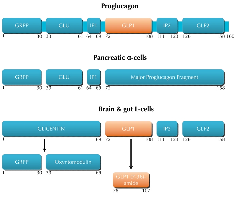
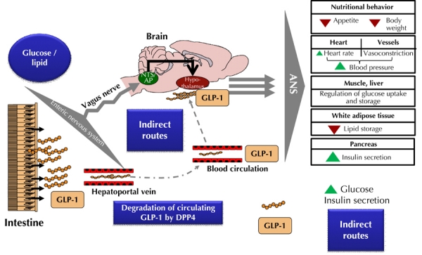
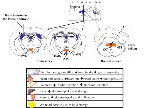
Glucagon-like peptide 1 (GLP-1) is a gut hormone which directly binds to the GLP-1 receptor located at the surface of the pancreatic β-cells to enhance glucose-induced insulin secretion. In addition to its pancreatic effects, GLP-1 can induce metabolic actions by interacting with its receptors expressed on nerve cells in the gut and the brain. GLP-1 can also be considered as a neuropeptide synthesized by neuronal cells in the brain stem that release the peptide directly into the hypothalamus. In this environment, GLP-1 is assumed to control numerous metabolic and cardiovascular functions such as insulin secretion, glucose production and utilization, and arterial blood flow. However, the exact roles of these two locations in the regulation of glucose homeostasis are not well understood. In this review, we highlight the latest experimental data supporting the role of the gut-brain and brain-periphery axes in the control of glucose homeostasis. We also focus our attention on the relevance of β-cell and brain cell targeting by gut GLP-1 for the regulation of glucose homeostasis. In addition to its action on β-cells, we find that understanding the physiological role of GLP-1 will help to develop GLP-1-based therapies to control glycemia in type 2 diabetes by triggering the gut-brain axis or the brain directly. This pleiotropic action of GLP-1 is an important concept that may help to explain the observation that, during their treatment, type 2 diabetic patients can be identified as 'responders' and 'non-responders'.
Copyright © by Lab & Life Press/SBDR
Figures
Similar articles
-
A role for the gut-to-brain GLP-1-dependent axis in the control of metabolism.Curr Opin Pharmacol. 2009 Dec;9(6):744-52. doi: 10.1016/j.coph.2009.09.003. Epub 2009 Sep 30. Curr Opin Pharmacol. 2009. PMID: 19800290 Review.
-
Glucagon-like Peptide-1 and the Central/Peripheral Nervous System: Crosstalk in Diabetes.Trends Endocrinol Metab. 2017 Feb;28(2):88-103. doi: 10.1016/j.tem.2016.10.001. Epub 2016 Oct 27. Trends Endocrinol Metab. 2017. PMID: 27871675 Review.
-
A Specific Gut Microbiota Dysbiosis of Type 2 Diabetic Mice Induces GLP-1 Resistance through an Enteric NO-Dependent and Gut-Brain Axis Mechanism.Cell Metab. 2017 May 2;25(5):1075-1090.e5. doi: 10.1016/j.cmet.2017.04.013. Cell Metab. 2017. PMID: 28467926
-
Gut incretin hormones in identical twins discordant for non-insulin-dependent diabetes mellitus (NIDDM)--evidence for decreased glucagon-like peptide 1 secretion during oral glucose ingestion in NIDDM twins.Eur J Endocrinol. 1996 Oct;135(4):425-32. doi: 10.1530/eje.0.1350425. Eur J Endocrinol. 1996. PMID: 8921824
-
Altered expression of uncoupling protein 2 in GLP-1-producing cells after chronic high glucose exposure: implications for the pathogenesis of diabetes mellitus.Am J Physiol Cell Physiol. 2016 Apr 1;310(7):C558-67. doi: 10.1152/ajpcell.00148.2015. Epub 2016 Jan 6. Am J Physiol Cell Physiol. 2016. PMID: 26739488
Cited by
-
An Update on the Effect of Incretin-Based Therapies on β-Cell Function and Mass.Diabetes Metab J. 2016 Apr;40(2):99-114. doi: 10.4093/dmj.2016.40.2.99. Diabetes Metab J. 2016. PMID: 27126881 Free PMC article. Review.
-
Regulation of Diabetes: a Therapeutic Strategy for Alzheimer's Disease?J Korean Med Sci. 2019 Dec 2;34(46):e297. doi: 10.3346/jkms.2019.34.e297. J Korean Med Sci. 2019. PMID: 31779058 Free PMC article. Review.
-
Bile Acid Signaling Pathways from the Enterohepatic Circulation to the Central Nervous System.Front Neurosci. 2017 Nov 7;11:617. doi: 10.3389/fnins.2017.00617. eCollection 2017. Front Neurosci. 2017. PMID: 29163019 Free PMC article. Review.
-
Sitagliptin and liraglutide reversed nigrostriatal degeneration of rodent brain in rotenone-induced Parkinson's disease.Inflammopharmacology. 2017 Jun;25(3):369-382. doi: 10.1007/s10787-017-0331-6. Epub 2017 Mar 4. Inflammopharmacology. 2017. PMID: 28258522
-
GLP-1 receptor agonists causing euglycemic ketoacidosis in patients without diabetes: a brief review.Int J Obes (Lond). 2025 Jun;49(6):977-979. doi: 10.1038/s41366-025-01749-x. Epub 2025 Mar 20. Int J Obes (Lond). 2025. PMID: 40114022 Review. No abstract available.
References
-
- Brown JC, Dryburgh JR, Ross SA, Dupre J. Identification and actions of gastric inhibitory polypeptide. Recent Prog Horm Res. 1975;31:487–532. - PubMed
-
- Dupre J, Beck JC. Stimulation of release of insulin by an extract of intestinal mucosa. Diabetes. 1966;15:555–559. - PubMed
-
- Elrick H, Stimmler L, Hlad CJ Jr, Arai Y. Plasma insulin response to oral and intravenous glucose administration. J Clin Endocrinol Metab. 1964;24:1076–1082. - PubMed
-
- McIntyre N, Holdsworth CD, Turner DS. Intestinal factors in the control of insulin secretion. J Clin Endocrinol Metab. 1965;25:1317–1324. - PubMed
Publication types
MeSH terms
Substances
LinkOut - more resources
Full Text Sources
Medical