

Haploinsufficiency of SGO1 results in deregulated centrosome dynamics, enhanced chromosomal instability and colon tumorigenesis
- PMID: 22262168
- PMCID: PMC3315092
- DOI: 10.4161/cc.11.3.18994
Haploinsufficiency of SGO1 results in deregulated centrosome dynamics, enhanced chromosomal instability and colon tumorigenesis
Abstract
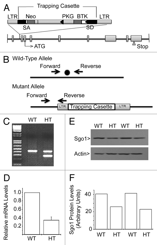
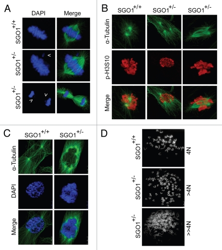
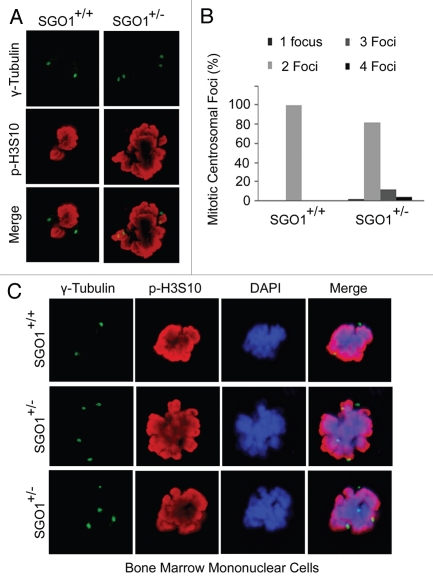
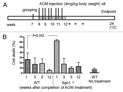
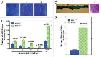
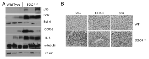
Chromosome instability (CIN) is found in 85% of colorectal cancers. Defects in mitotic processes are implicated in high CIN and may be critical events in colorectal tumorigenesis. Shugoshin-1 (SGO1) aids in the maintenance of chromosome cohesion and prevents premature chromosome separation and CIN. In addition, integrity of the centrosome may be compromised due to the deficiency of Cohesin and Sgo1 through the disengagement of centrioles. We report here the generation and characterization of SGO1-mutant mice and show that haploinsufficiency of SGO1 leads to enhanced colonic tumorigenesis. Complete disruption of SGO1 results in embryonic lethality, whereas SGO1+/- mice are viable and fertile. Haploinsufficiency of SGO1 results in genomic instability manifested as missegregation of chromosomes and formation of extra centrosomal foci in both murine embryonic fibroblasts and adult bone marrow cells. Enhanced CIN observed in SGO1-deficient mice resulted in an increase in formation of aberrant crypt foci (ACF) and accelerated development of tumors after exposure to azoxymethane (AOM), a colon carcinogen. Together, these results suggest that haploinsufficiency of SGO1 causes enhanced CIN, colonic preneoplastic lesions and tumorigenesis in mice. SGO1 is essential for the suppression of CIN and tumor formation.
Figures
Comment in
-
Sgo1 as a "guardian spirit" for preventing colon tumorigenesis.Cell Cycle. 2012 Feb 15;11(4):649. doi: 10.4161/cc.11.4.19360. Cell Cycle. 2012. PMID: 22374668 Free PMC article. No abstract available.
-
Sgo1 as a guardian of chromosome stability.Cell Cycle. 2012 Feb 15;11(4):650-1. doi: 10.4161/cc.11.4.19361. Cell Cycle. 2012. PMID: 22374669 No abstract available.
Similar articles
-
Antagonizing pathways leading to differential dynamics in colon carcinogenesis in Shugoshin1 (Sgo1)-haploinsufficient chromosome instability model.Mol Carcinog. 2016 May;55(5):600-10. doi: 10.1002/mc.22306. Epub 2015 Mar 14. Mol Carcinog. 2016. PMID: 25773652 Free PMC article.
-
Sgo1 as a "guardian spirit" for preventing colon tumorigenesis.Cell Cycle. 2012 Feb 15;11(4):649. doi: 10.4161/cc.11.4.19360. Cell Cycle. 2012. PMID: 22374668 Free PMC article. No abstract available.
-
Tumor-promoting/progressing role of additional chromosome instability in hepatic carcinogenesis in Sgo1 (Shugoshin 1) haploinsufficient mice.Carcinogenesis. 2015 Apr;36(4):429-40. doi: 10.1093/carcin/bgv011. Epub 2015 Mar 4. Carcinogenesis. 2015. PMID: 25740822 Free PMC article.
-
Critical role of mitosis in spontaneous late-onset Alzheimer's disease; from a Shugoshin 1 cohesinopathy mouse model.Cell Cycle. 2018;17(19-20):2321-2334. doi: 10.1080/15384101.2018.1515554. Epub 2018 Sep 20. Cell Cycle. 2018. PMID: 30231670 Free PMC article. Review.
-
The expanding phenotypes of cohesinopathies: one ring to rule them all!Cell Cycle. 2019 Nov;18(21):2828-2848. doi: 10.1080/15384101.2019.1658476. Epub 2019 Sep 13. Cell Cycle. 2019. PMID: 31516082 Free PMC article. Review.
Cited by
-
Chromosome Instability and Mosaic Aneuploidy in Neurodegenerative and Neurodevelopmental Disorders.Front Genet. 2019 Nov 7;10:1092. doi: 10.3389/fgene.2019.01092. eCollection 2019. Front Genet. 2019. PMID: 31788001 Free PMC article.
-
SGO1 maintains bovine meiotic and mitotic centromeric cohesions of sister chromatids and directly affects embryo development.PLoS One. 2013 Sep 3;8(9):e73636. doi: 10.1371/journal.pone.0073636. eCollection 2013. PLoS One. 2013. PMID: 24019931 Free PMC article.
-
GSK3-ARC/Arg3.1 and GSK3-Wnt signaling axes trigger amyloid-β accumulation and neuroinflammation in middle-aged Shugoshin 1 mice.Aging Cell. 2020 Oct;19(10):e13221. doi: 10.1111/acel.13221. Epub 2020 Aug 28. Aging Cell. 2020. PMID: 32857910 Free PMC article.
-
SGO1 induces proliferation and metastasis of prostate cancer through AKT-mediated signaling pathway.Am J Cancer Res. 2019 Dec 1;9(12):2693-2705. eCollection 2019. Am J Cancer Res. 2019. PMID: 31911855 Free PMC article.
-
Chromosome instability and aneuploidy as context-dependent activators or inhibitors of antitumor immunity.Front Immunol. 2022 Jul 15;13:895961. doi: 10.3389/fimmu.2022.895961. eCollection 2022. Front Immunol. 2022. PMID: 36003402 Free PMC article. Review.
References
Publication types
MeSH terms
Substances
Grants and funding
LinkOut - more resources
Full Text Sources
Molecular Biology Databases