. 2012 Feb 1;11(3):524-31.
doi: 10.4161/cc.11.3.19033. Epub 2012 Feb 1.

Cdc6 is required for meiotic spindle assembly in Xenopus oocytes

Affiliations

Cdc6 is required for meiotic spindle assembly in Xenopus oocytes

Yadushyla Narasimhachar et al. Cell Cycle. .

Abstract

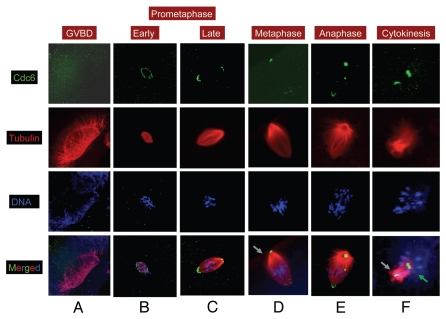
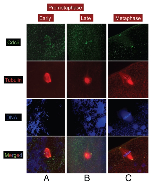
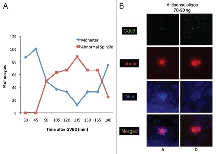
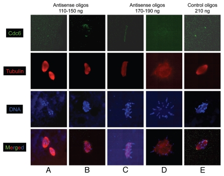
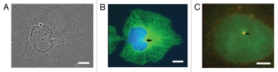
During the maturation of Xenopus oocytes, Cdc6 expression is necessary to establish replication competence to support early embryonic DNA replication. However, Cdc6 is expressed before the completion of MI, at a time when its function as a replication factor is not required, suggesting additional roles for Cdc6 in meiosis. Confocal immunofluorescence microscopy revealed that Cdc6 protein was distributed around the spindle precursor at the time of germinal vesicle breakdown (GVBD), and localized to the margin of the nascent spindle early in prometaphase. Cdc6 subsequently localized to spindle poles in late prometaphase, where it remained until metaphase arrest. Microinjection of antisense oligonucleotides specific for Cdc6 mRNA disrupted spindle assembly, resulting in defects including delayed spindle assembly, misoriented and unattached anaphase spindles, monasters, multiple spindles, microtubule aggregates associated with condensed chromosomes, or the absence of recognizable spindle-like structures, depending on the level of residual Cdc6 expression. Furthermore, Cdc6 co-localized with γ-tubulin in centrosomes during interphase in all somatic cells analyzed, and associated with spindle poles in mitotic COS cells. Our data suggest a role for Cdc6 in spindle formation in addition to its role as a DNA replication factor.


Figures

Figure 1
Figure 1
Cdc6 is localized to spindle poles in MI. Stage VI oocytes were treated with progesterone to induce maturation, and samples were collected at various time and processed for immunofluorescence. Oocytes were stained for α tubulin (red), Cdc6 (green) and DNA (blue). Grey arrow in (D) indicates the animal cortex. Grey arrow in panel f indicates the first polar body and green arrow indicates the second meiotic spindle precursor. Image scale: 140 µm (A), 80 µm (B), 40–50 µm (CµF).
Figure 2
Figure 2
Cdc6 localizes to spindle poles in MII. Stage VI oocytes were treated with progesterone to induce maturation, and samples were collected at various time and processed for immunofluorescence. Samples were stained for α tubulin (red), Cdc6 (green) and DNA (blue). Image scale: 90 µm.
Figure 3
Figure 3
Inhibition of Cdc6 during maturation leads to delayed spindle assembly. Stage VI oocytes were injected with 70–90 ng of Cdc6 antisense or control non-specific oligonucleotides and treated with progesterone to induce maturation, and samples were fixed at various times and stained for α tubulin (red), Cdc6 (green) and DNA (blue). (A) Plot represents the % of oocytes with monasters or abnormal spindles (delayed spindles with abnormal or normal morphology) at indicated time during maturation. [B(a)] Monaster formation in Cdc6 antisense injected oocyte during MI (refer to Fig. 1C for control/normal spindle at this time). [B(b)] An unattached anaphase/telophase spindle seen at 135 min post-GVBD (refer to Fig. 1E and F for control/normal spindle phenotype at this time). Image scale: 70 µm.
Figure 4
Figure 4
Inhibition of Cdc6 during maturation causes abnormal spindle assembly. Stage VI oocytes were injected with various concentrations of Cdc6 antisense or control non-specific oligonucleotides, treated with progesterone to induce maturation and samples were collected during early MI (between 30–60 min post-GVBD) and stained for α tubulin (red), Cdc6 (green) and DNA (blue). (A and B) At 110–150 ng of antisense injection, multiple spindles, tripolar spindles were noted. (C and D) At 170–190 ng of antisense injection, elongated, distorted spindle-like structure or tubulin mass attached with condensed chromosomes were seen. (E) Control non-specific oligonucleotides injected (210 ng) oocytes had normal prometaphase MI spindle. Image scale: 65–70 µm.
Figure 5
Figure 5
Cdc6 co-localizes with γ-tubulin at interphase centrosomes of mammalian cells. Affinity-purified antibodies to Cdc6 protein stain puncta at the perinuclear focus of microtubule staining (the centrosome) in a variety of mammalian cell lines, including HeLa cells [shown here in (B)]. At either lower (B) or higher (C) magnification, the colocalization of Cdc6 with one (or sometimes both) puncta of centrosomes (stained with antibodies to gamma tubulin) is observed. Cdc6 stained red; γ-tubulin (C) or α-tubulin (B) stained green, and DNA stained blue with Hoechst. (A) is the phase contrast image of HeLa cells. Scale bars in (A and B = 5 micron) and (C = 1 micron).

Similar articles

Cited by

References

    1. Sutton WS. On the morphology of the chromosome group in Brachystola Magna. Biol Bull. 1902;4:24–39. doi: 10.2307/1535510. - DOI
    1. Sutton WS. The chromosomes in heredity. Biological Bulletin. Marine Biological Laboratory. 1903;4:231–251. doi: 10.2307/1535741. - DOI
    1. Farmer JB, Moore JES. On the maiotic phase (reduction divisions) in animals and plants. Q J Microsc Sci. 1905;48:489–557.
    1. Gurdon JB. On the origin and persistence of a cytoplasmic state inducing nuclear DNA synthesis in frogs' eggs. Proc Natl Acad Sci USA. 1967;58:545–552. doi: 10.1073/pnas.58.2.545. - DOI - PMC - PubMed
    1. Whitmire E, Khan B, Coué M. Cdc6 synthesis regulates replication competence in Xenopus oocytes. Nature. 2002;419:722–725. doi: 10.1038/nature01032. - DOI - PubMed

Publication types

MeSH terms

LinkOut - more resources