. 2012 Mar;23(6):1080-94.
doi: 10.1091/mbc.E11-07-0588. Epub 2012 Jan 19.

Ezrin regulates microvillus morphogenesis by promoting distinct activities of Eps8 proteins

Affiliations

Ezrin regulates microvillus morphogenesis by promoting distinct activities of Eps8 proteins

Ingrid Zwaenepoel et al. Mol Biol Cell. 2012 Mar.

Abstract

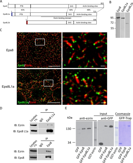
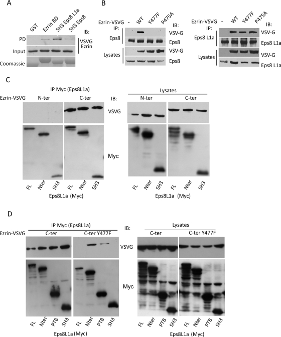
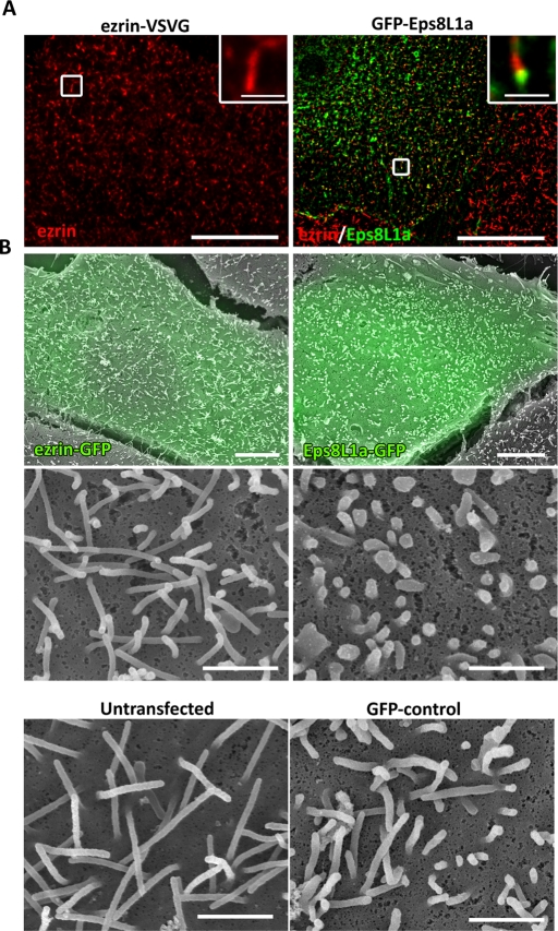
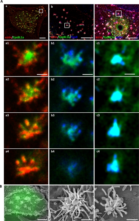
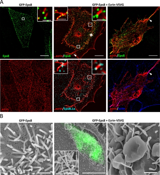
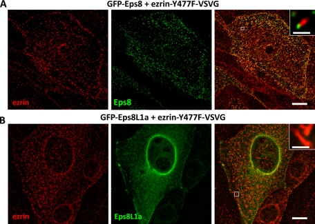
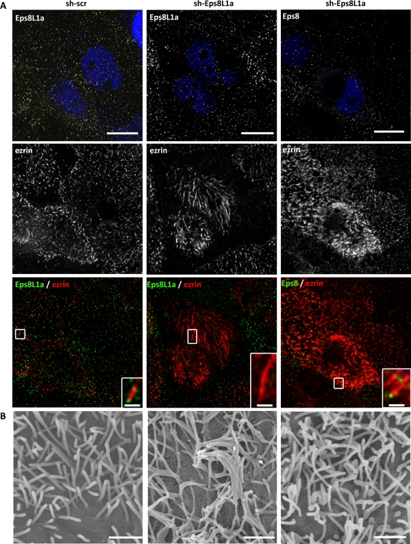
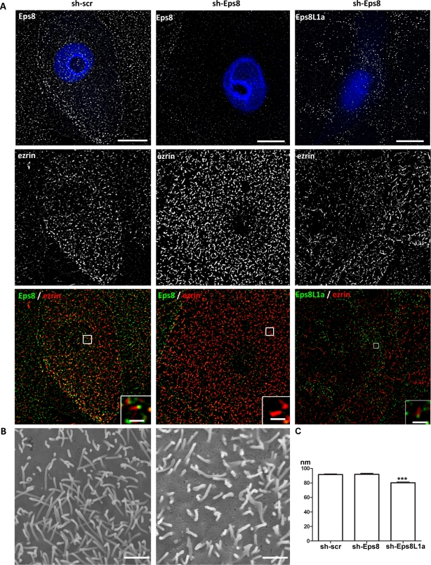
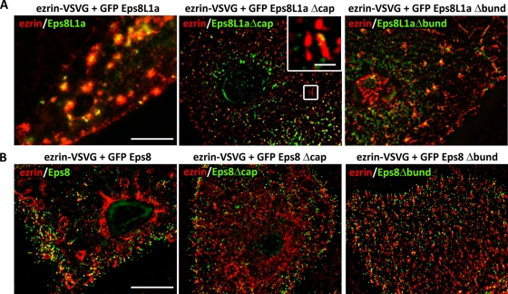
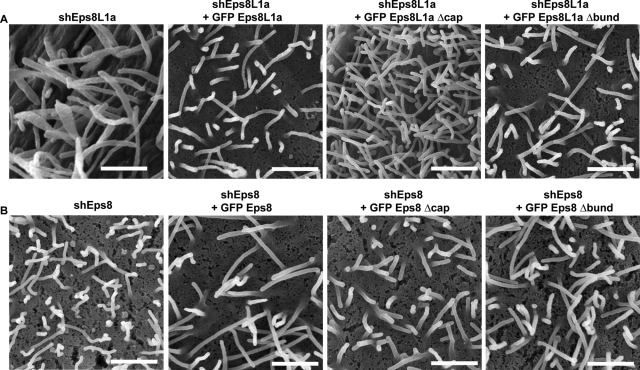
The mechanisms that regulate actin filament polymerization resulting in the morphogenesis of the brush border microvilli in epithelial cells remain unknown. Eps8, the prototype of a family of proteins capable of capping and bundling actin filaments, has been shown to bundle the microvillar actin filaments. We report that Eps8L1a, a member of the Eps8 family and a novel ezrin-interacting partner, controls microvillus length through its capping activity. Depletion of Eps8L1a leads to the formation of long microvilli, whereas its overexpression has the opposite effect. We demonstrate that ezrin differentially modulates the actin-capping and -bundling activities of Eps8 and Eps8L1a during microvillus assembly. Coexpression of ezrin with Eps8 promotes the formation of membrane ruffles and tufts of microvilli, whereas expression of ezrin and Eps8L1a induces the clustering of actin-containing structures at the cell surface. These distinct morphological changes are neither observed when a mutant of ezrin defective in its binding to Eps8/Eps8L1a is coexpressed with Eps8 or Eps8L1a nor observed when ezrin is expressed with mutants of Eps8 or Eps8L1a defective in the actin-bundling or -capping activities, respectively. Our data show a synergistic effect of ezrin and Eps8 proteins in the assembly and organization of actin microvillar filaments.


Figures

FIGURE 1:
FIGURE 1:
Eps8 and Eps8L1a are components of the brush border microvilli. (A) Schematic representation of the structure of Eps8 and Eps8L1a and b isoforms. The percentage of identity of Eps8 domains with that of Eps8L1a and b is indicated. The position of the peptides used to generate antibodies against Eps8L1a and b is represented by the blue and red boxes, respectively. The region of Eps8L1a and b found to interact with ezrin in the two-hybrid screen is underlined. (B) Western blot was performed on LLC-PK1 cell extracts using anti-Eps8, anti-Eps8L1a, and anti-Eps8L1b antibodies. (C) Localization of Eps8 proteins in LLC-PK1 cells. Double immunofluorescence was performed with an anti-ezrin antibody (red) and either anti-Eps8 (top) or Eps8L1a (bottom) antibodies (green). Images were taken with a three-dimensional wide-field optical sectioning microscope. The images on the left correspond to a maximum-intensity Z projection, performed after deconvolution, of 10 slices starting from the top of the cells. Bar, 10 μm. Microvilli in the rectangles are shown at high magnification on the right. Bar, 1 μm. (D) Immunoprecipitation was performed with control immunoglobulin G (IgG) or anti-Eps8L1a (top) and anti-Eps8 (bottom) antibodies, respectively, from LLC-PK1 cell lysates. Thirty percent of the immunoprecipitates were loaded on the gel. The Western blot performed with an anti-ezrin antibody indicates that ezrin coimmunoprecipitates with Eps8 and Eps8L1a. (E) In vitro interaction of GFP-Eps8 or GFP-Eps8L1a with ezrin. GFP, GFP-Eps8, or GFP- Eps8L1a bound to GFP-Trap_A beads were incubated with purified GST-ezrin. Left, the immunoblot performed with the anti-ezrin antibody shows that ezrin interacts with Eps8 and Eps8L1a but not with GFP alone. One-fifth of the beads were loaded on the gel. Middle, 1/300 and 1/25 of purified GST-ezrin and GFP proteins used for the interaction were loaded on the gel and analyzed with anti-ezrin and anti-GFP antibodies, respectively. Right, Coomassie staining of purified proteins.
FIGURE 2:
FIGURE 2:
Mapping of interaction sites between ezrin and Eps8 proteins. (A) Immobilized GST or GST fused to ezrin-binding domain on Eps8L1a (BD), the SH3 domain of Eps8L1a, and the SH3 domain of Eps8 were incubated with lysates of LLC-PK1 cells expressing ezrin-VSVG. Western blot was performed with an anti-VSVG antibody. Bottom, the Coomassie staining of GST-fused proteins. (B) Left, immunoprecipitation performed with an anti-Eps8 antibody on lysates of LLC-PK1 cells stably transfected with the empty vector (–) or with cDNA coding ezrin wild type (WT), ezrinY477F, or P475A tagged with VSVG. Immunoblots were performed with an anti-VSVG antibody to detect ezrin. Eps8 interacts only with wild-type ezrin. Right, the immunoprecipitation was performed as before, but with an anti-Eps8L1a antibody. Eps8L1a interacts with wild-type ezrin, as well as with ezrin Y477F and ezrin P475A. (C) 293T cells were cotransfected with cDNA coding the VSVG-tagged carboxy-terminal (C-ter) or amino-terminal (N-ter) domains of ezrin and Myc-tagged full-length Eps8L1a (FL) or its various domains. Left, immunoprecipitation performed with an anti-Myc antibody, followed by an immunoblot on immunoprecipitated proteins with anti-Myc and anti-VSVG antibodies. Right, cell lysates analyzed with anti-VSVG and anti- Myc antibodies. (D) 293T cells were cotransfected with cDNA coding the VSVG tagged carboxy-terminal domain of ezrin carrying or not the mutation Y477F and Myc-tagged full-length Eps8L1a or its various domains. Left, immunoprecipitation performed with an anti-myc antibody, followed by Western blots with anti-VSVG and anti-Myc antibodies. Right, cell lysates analyzed with anti-VSVG and anti-Myc antibodies. The introduction of the Y477F mutation in the carboxy-terminal domain of ezrin abolishes the interaction of Eps8L1a SH3 domain with ezrin.
FIGURE 3:
FIGURE 3:
Expression of Eps8L1a blocks microvillus elongation. (A) Immunofluorescence was performed on LLC-PK1 cells expressing ezrin-VSVG (left) or GFP-Eps8L1a (right). Ezrin (in red) was detected with an anti-VSVG antibody. Images were taken as in Figure 1B and correspond to a maximum-intensity Z-projection performed after deconvolution. Insets show the localization of ezrin along the microvilli (left) and Eps8L1a at the tip (right). Bar, 10 μm; insets, 1 μm. (B) CL-SEM was performed on cells expressing ezrin-GFP or GFP-Eps8L1a. Transfected cells (in green) were tracked by fluorescence and subsequently analyzed by scanning electron microscopy. The size of the microvilli is considerably reduced in cells expressing Eps8L1a as compared with that of cells expressing ezrin-GFP or of untransfected cells (bottom). Bars, first row,10 μm; others, 1 μm.
FIGURE 4:
FIGURE 4:
Coexpression of ezrin and Eps8L1a induces microvillar clustering at the apical surface of LLC-PK1 cells. LLC-PK1 cells were cotransfected with plasmids coding for ezrin-VSVG and GFP-Eps8L1a. (A) Immunofluorescence was performed with an anti-VSVG antibody to detect ezrin (a, b, c, a1–a4; red) and an anti-Eps8 antibody (b, b1–b4; blue). Actin was detected with phalloidin (c, c1–c4; blue). Images correspond to a maximum-intensity Z-projection performed after deconvolution. Bottom, four successive slices of the clusters in the insets from a–c are shown from the bottom (a1, b1, c1) to the top of the structure (a4, b4, c4). Eps8L1a is mainly localized in the enlarged membrane structures (see scanning electron microscopy) and ezrin in the microvilli that originate from them. Bars, a–c, 10 μm; a1–c4, 1 μm. (B) CL-SEM. Low (left) and high (middle and right) magnification of the surface of LLC-PK1 cells expressing ezrin-VSVG and GFP-Eps8L1a. Clusters from two different cells are shown at high magnification. Transfected cells are monitored by fluorescence through GFP fluorescence. Bars, left, 10 μm; middle and right, 1 μm.
FIGURE 5:
FIGURE 5:
Coexpression of ezrin and Eps8 induces the formation of membrane ruffles and tufts of microvilli at the apical surface of LLC-PK1 cells. (A) Immunofluorescence of cells expressing GFP-Eps8 alone or GFP-Eps8 and ezrin-VSVG. Images correspond to a maximum-intensity Z-projection performed after deconvolution. Dorsal circular ruffles, arc-shaped ridges covered with abundant microvilli (asterisk, middle), and actin bundles (arrow, right) are observed in cells expressing both proteins. Insets (right of the middle) show the localization of Eps8 and Eps8L1a at the tip of the microvilli. A punctate staining of Eps8 and Eps8L1a is observed in membrane ruffles (left insets, middle). Bars, 10 μm; inset, 1 μm. (B) CL-SEM. Left, microvilli of cells overexpressing GFP-Eps8. Middle and right, low and high magnification of the surface of cells expressing ezrin and GFP-Eps8. Extensive dorsal ruffles are observed. The inset shows the tufts of microvilli observed at the edge of the cells. Bars, left, 1 μm; middle, 10 μm (inset, 1 μm); right, 1 μm.
FIGURE 6:
FIGURE 6:
Ezrin Y477F fails to promote Eps8 protein activities. Immunofluorescence was performed on LLC-PK1 cells coexpressing ezrin Y477F and (A) GFP-Eps8 or (B) GFP-Eps8L1a with an anti-VSVG antibody to detect ezrin (red). No morphological changes are observed in cells expressing both proteins. (A) Eps8 remains localized at the tip of the microvilli (inset, right). (B) Eps8L1a is diffuse in the cytoplasm and absent from the tip of the microvilli (inset, right). Bars, 10 μm; inset, 1 μm.
FIGURE 7:
FIGURE 7:
Depletion of Eps8L1a induces the formation of long microvilli in LLC-PK1 cells. Cells transfected with plasmids coding the reporter gene GFP and shRNA, nontargeting (scr; left) or targeting Eps8L1a (middle and right) were analyzed by immunofluorescence. Transfected cells were monitored through the expression of GFP (artificially rendered in blue). Immunofluorescence was performed with anti-ezrin (red) and anti-Eps8L1a and anti-Eps8 (green) antibodies. Images correspond to a maximum-intensity Z-projection performed after deconvolution. Eps8 is still localized at the tip of the microvilli (right). Bars, 10 μm; inset, 1 μm. (B) CL-SEM shows microvilli of cells expressing scramble (left) or Eps8L1a shRNA. Bar, 1 μm. Both immunofluorescence and CL-SEM indicate that depletion of Eps8L1a leads to the formation of long microvilli.
FIGURE 8:
FIGURE 8:
LLC-PK1 cells depleted for Eps8 display irregular microvilli at their apical surface. (A) Immunofluorescence and (B) CL-SEM were performed on cells expressing shRNA scr (left) or targeting Eps8 (middle and right). Transfected cells were detected through the expression of GFP (artificially rendered in blue). Eps8L1a localizes to the tip of the microvilli (right). Bars, 10 μm; inset, 1 μm. (B) CL-SEM indicates that cells depleted for Eps8 display shorter and more irregular microvilli. Bar, 1 μm. (C) Graph showing the width of the microvilli determined from CL-SEM images of cells transfected with Scr, Eps8, or Eps8L1a psiRNA. The values in the histogram are means ± SE. Statistical significance was determined by one-way analysis of variance with a Dunnett multiple comparison test. The width (nm) of the microvilli in cells depleted of Eps8L1a is significantly thinner than in cells transfected with nontargeting or Eps8-targeting shRNA, with p < 0.005.
FIGURE 9:
FIGURE 9:
Actin-capping and -bundling activities of Eps8L1a and Eps8, respectively, in microvillus assembly. (A) LLC-PK1 cells were transfected with plasmids coding ezrin and GFP-Eps8L1a (left), ezrin and Eps8L1a mutated in its capping site (Eps8L1a∆cap, middle), or ezrin and Eps8L1a mutated in its actin-bundling site (Eps8L1a∆bund, right). Clusters are observed in cells expressing ezrin with either Eps8L1 wild type or Eps8L1aΔbund but not with Eps8L1aΔcap. Inset in the middle shows that Eps8L1a∆cap is localized at the microvilli side and rarely at their tip as observed with Eps8L1a. Bars, 10 μm; inset, 1 μm. (B) LLC-PK1 cells were transfected with plasmids coding ezrin and GFP-Eps8 (left), ezrin and GFP-Eps8 mutated in its capping site (middle, Eps8Δcap), or ezrin and Eps8 mutated in its actin-bundling site (right, Eps8Δbund). Circular dorsal ruffles are observed on the surface of the cells coexpressing ezrin and Eps8 or Eps8Δcap but not in cells expressing ezrin and Eps8Δbund. Bars, 10 μm; inset, 1 μm.
FIGURE 10:
FIGURE 10:
The defects caused by depletion of either Eps8 or Eps8L1a can be reverted by expressing wild-type Eps8 or Eps8L1a, respectively, but not their mutant forms. (A) LLC-PK1 cells expressing shRNA targeting Eps8L1a were transfected with human GFP-Eps8L1a wild type, GFP-Eps8L1aΔcap, or GFP-Eps8L1aΔbund. Long microvilli are observed in cells depleted for Eps8L1a with or without expression of Eps8L1aΔcap, whereas shorter microvilli are observed in cells expressing the shRNA targeting Eps8L1a and Eps8L1a wild type or Eps8L1aΔbund. (B) LLC-PK1 cells expressing shRNA targeting Eps8 were transfected with human GFP-Eps8 wild type, GFP-Eps8Δcap, or GFP-Eps8Δbund. Cells expressing shRNA targeting Eps8 or cells expressing shRNA targeting Eps8 and GFP-Eps8Δbund display irregular, bent microvilli. In contrast, cells expressing shRNA targeting Eps8 and either wild-type GFP-Eps8 or GFP-Eps8 Δcap show stiff microvilli. Bar, 10 μm.

Similar articles

Cited by

References

    1. Abramoff MD, Magelhaes PJ, Ram SJ. Image processing with ImageJ. Biophotonics Int. 2004;11:36–42.
    1. Algrain M, Arpin M, Louvard D. Wizardry at the cell cortex. Cur Biol. 1993;3:451–454. - PubMed
    1. Andreoli C, Martin M, Leborgne R, Reggio H, Mangeat P. Ezrin has properties to self-associate at the plasma membrane. J Cell Sci. 1994;107:2509–2521. - PubMed
    1. Arpin M, Chirivino D, Naba A, Zwaenepoel I. Emerging role for ERM proteins in cell adhesion and migration. Cell Adhes Migration. 2011;5:199–206. - PMC - PubMed
    1. Bretscher A. Rapid phosphorylation and reorganization of ezrin and spectrin accompany morphological changes induced in A-431 cells by epidermal growth factor. J Cell Biol. 1989;108:921–930. - PMC - PubMed

Publication types

LinkOut - more resources