. 2012 Apr 27;86(4):130.
doi: 10.1095/biolreprod.111.094870. Print 2012 Apr.

Patterns of gene expression in the bovine corpus luteum following repeated intrauterine infusions of low doses of prostaglandin F2alpha

Affiliations

Patterns of gene expression in the bovine corpus luteum following repeated intrauterine infusions of low doses of prostaglandin F2alpha

Mehmet O Atli et al. Biol Reprod. .

Abstract

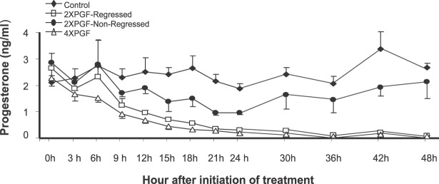
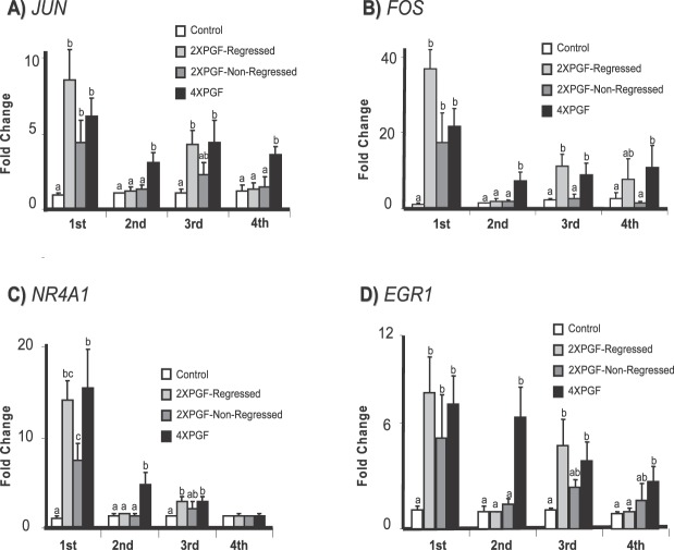
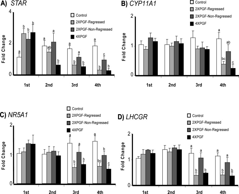
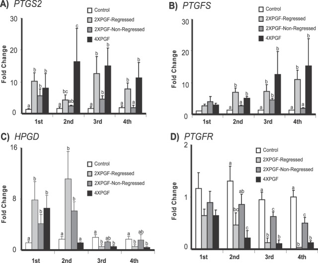
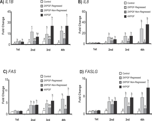
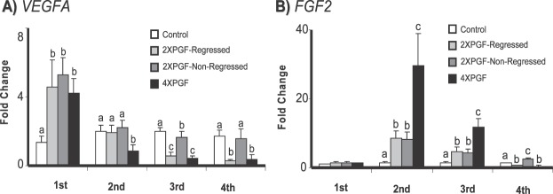
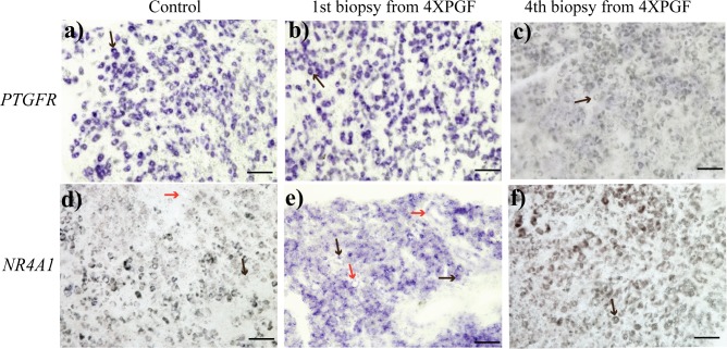
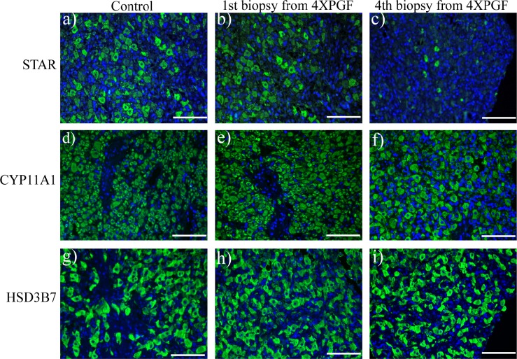
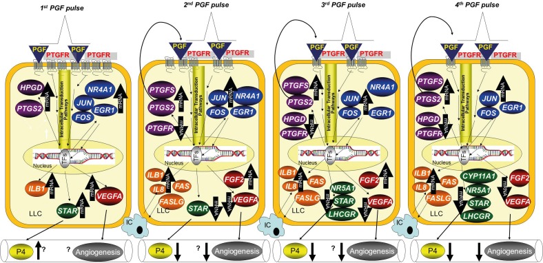
Natural luteolysis involves multiple pulses of prostaglandin F2alpha (PGF) released by the nonpregnant uterus. This study investigated expression of 18 genes from five distinct pathways, following multiple low-dose pulses of PGF. Cows on Day 9 of the estrous cycle received four intrauterine infusions of 0.25 ml of phosphate-buffered saline (PBS) or PGF (0.5 mg of PGF in 0.25 ml of PBS) at 6-h intervals. A luteal biopsy sample was collected 30 min after each PBS or PGF infusion. There were four treatment groups: Control (n = 5; 4 PBS infusions), 4XPGF (4 PGF infusions; n = 5), 2XPGF-non-regressed (2 PGF infusions; n = 5; PGF-PBS-PGF-PBS; no regression after treatments), and 2XPGF-regressed (PGF-PBS-PGF-PBS; regression after treatments; n = 5). As expected, the first PGF pulse increased mRNA for the immediate early genes JUN, FOS, NR4A1, and EGR1 but unexpectedly also increased mRNA for steroidogenic (STAR) and angiogenic (VEGFA) pathways. The second PGF pulse induced immediate early genes and genes related to immune system activation (IL1B, FAS, FASLG, IL8). However, mRNA for VEGFA and STAR were decreased by the second PGF infusion. After the third and fourth PGF pulses, a distinctly luteolytic pattern of gene expression was evident, with inhibition of steroidogenic and angiogenic pathways, whereas, there was induction of pathways for immune system activation and production of PGF. The pattern of PGF-induced gene expression was similar in corpus luteum not destined for luteolysis (2X-non-regressed) after the first PGF pulse but was very distinct after the second PGF pulse. Thus, although the initial PGF pulse induced mRNA for many pathways, the second and later pulses of PGF appear to have set the distinct pattern of gene expression that result in luteolysis.


Figures

FIG. 1
FIG. 1
Effect of intrauterine PGF or PBS treatment on circulating P4 concentration in cows. Data are presented as means ± SEM.
FIG. 2
FIG. 2
Effect of intrauterine PGF or PBS treatment on steady-state mRNA concentrations for A) JUN (jun proto-oncogene), B) FOS (FBJ murine osteosarcoma viral oncogene homolog), C) NR4A1 (nuclear receptor subfamily 4, group A, member 1), and D) EGR1 (early growth response 1). Data are shown as fold changes ± SEM. Columns with different letters (a, b, c) at each biopsy indicate differences; P < 0.05. First, second, third, and fourth indicate biopsy times.
FIG. 3
FIG. 3
Effect of intrauterine PGF or PBS treatment on steady-state mRNA concentrations for A) STAR (steroidogenic acute regulatory protein), B) CYP11A1 (cytochrome P450, family 11, subfamily A, polypeptide 1), C) NR5A1 (nuclear receptor subfamily 5, group A, member 1), and D) LHCGR (luteinizing hormone/choriogonadotropin receptor). Data are shown as fold changes ± SEM. Columns with different letters (a, b, c) at each biopsy indicate differences; P < 0.05. First, second, third, and fourth indicate biopsy times.
FIG. 4
FIG. 4
Effect of intrauterine PGF or PBS treatment on steady-state mRNA concentrations for A) PTGS2 (prostaglandin-endoperoxide synthase 2), B) PTGFS (prostaglandin F synthase), C) HPGD (hydroxyprostaglandin dehydrogenase 15-[NAD]), and D) PTGFR (prostaglandin F receptor). Data are shown as fold changes ± SEM. Columns with different letters (a, b, c) at each biopsy indicate differences, P < 0.05. First, second, third, and fourth indicate biopsy times.
FIG. 5
FIG. 5
Effect of intrauterine PGF or PBS treatment on steady-state mRNA concentrations for A) IL1B (interleukin 1, beta), B) IL8 (interleukin 8), C) FAS (Fas, TNF receptor superfamily, member 6), D) FASLG (Fas ligand). Data are shown as fold changes ± SEM. Columns with different letters (a, b, c) at each biopsy indicate differences, P < 0.05. First, second, third, and fourth indicate biopsy times.
FIG. 6
FIG. 6
Effect of intrauterine PGF or PBS treatment on steady-state mRNA concentrations for A) VEGFA (vascular endothelial growth factor A), B) FGF2 (fibroblast growth factor 2). Data are shown as fold changes ± SEM. Columns with different letters (a, b, c) at each biopsy indicate differences, P < 0.05. First, second, third, and fourth indicate biopsy times.
FIG. 7
FIG. 7
Representative images of in situ hybridization staining for PTGFR (prostaglandin F receptor, a, b, c) and NR4A1 (nuclear receptor subfamily 4, group A, member 1, d, e, f) in bovine CL in control and at first, and fourth biopsies from 4XPGF group. Large cells (black arrow) and vascular endothelial cells (red arrow) are shown. Bars = 100 μm.
FIG. 8
FIG. 8
Representative images of immunofluorescent staining for STAR (steroidogenic acute regulatory protein a, b, c), CYP11A1 (cytochrome P450, family 11, subfamily A, polypeptide 1, d, e, f), and HSD3B7 (hydroxy-delta-5-steroid dehydrogenase, 3 beta- and steroid delta-isomerase 7, g, h, i) in bovine CL at control (30 min after saline), and at first and fourth biopsies from 4XPGF group. Labeled tissue sections were counterstained with 4′, 6-diamidino-2-phenylindole dilactate (blue staining). Bars = 100 μm.
FIG. 9
FIG. 9
Schematic model shows changes in mRNAs following the four PGF pulses associated with luteolysis. Following the first PGF pulse, there were increases in mRNA for specific genes in all five pathways that were examined: immediate early genes (blue), PGF-related genes (purple), immune-related genes (orange), steroidogenic genes (green), and angiogenesis genes (red). Increases were not specific for luteal regression as similar increases were observed in CL that subsequently underwent or did not undergo regression. The second PGF pulse also increased the immediate early genes, but this pulse induced decreases in VEGFA and STAR. These changes were specific for the second PGF pulse, as they did not happen in CL that were only exposed to a single PGF pulse. After the third PGF pulse, there were dramatic changes in mRNA, consistent with a course that would result in luteolysis. Most of these changes did not happen in CL that were not destined for luteolysis, even though they also received the second PGF pulse at 30 min prior to the third biopsy. The fourth biopsy continued the luteolytic cascade with increases or decreases in essentially all genes in a luteolytic direction that was specific for CL that were undergoing the full luteolytic cascade. Names of genes are defined in the text. An up arrow (↑) indicates up-regulation; a down arrow (↓) indicates down-regulation; IC, immune cell; LLC, large luteal cells; TFs, transcription factors; P4, progesterone. The question marks indicate possible or unknown outcomes such as increased P4 following STAR or changes in angiogenesis after changes in VEGFA or FGF2.

Comment in

References

    1. O'Shea JD, Rodgers RJ, D'Occhio MJ. Cellular composition of the cyclic corpus luteum of the cow. J Reprod Fertil 1989; 85: 483 487 - PubMed
    1. Wiltbank MC. Cell types and hormonal mechanisms associated with mid-cycle corpus luteum function. J Anim Sci 1994; 72: 1873 1883 - PubMed
    1. Niswender GD, Juengel JL, McGuire WJ, Belfiore CJ, Wiltbank MC. Luteal function: the estrous cycle and early pregnancy. Biol Reprod 1994; 50: 239 247 - PubMed
    1. Niswender GD, Juengel JL, Silva PJ, Rollyson MK, McIntush EW. Mechanisms controlling the function and life span of the corpus luteum. Physiol Rev 2000; 80: 1 29 - PubMed
    1. Schams D, Berisha B. Regulation of corpus luteum function in cattle—an overview. Reprod Domest Anim 2004; 39: 241 251 - PubMed

Publication types