. 2012 Mar 16;287(12):9429-40.
doi: 10.1074/jbc.M111.304329. Epub 2012 Jan 19.

Constitutive clathrin-mediated endocytosis of CTLA-4 persists during T cell activation

Affiliations

Constitutive clathrin-mediated endocytosis of CTLA-4 persists during T cell activation

Omar S Qureshi et al. J Biol Chem. .

Abstract

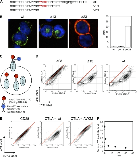
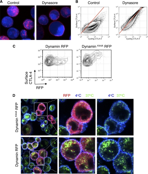
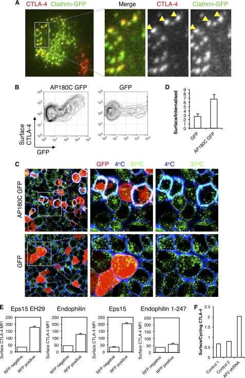
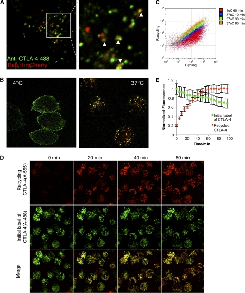
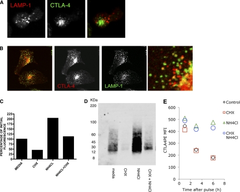
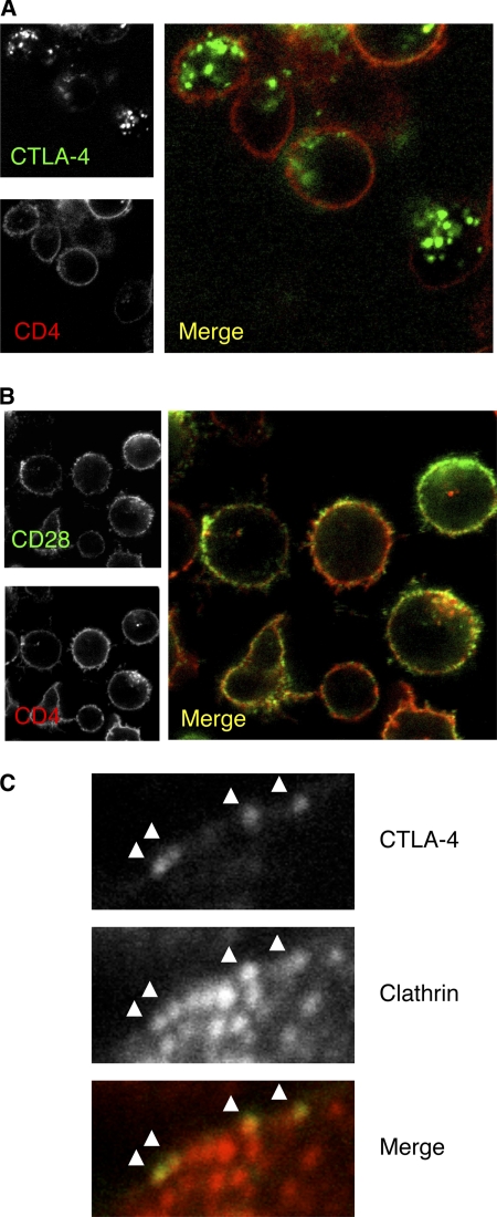
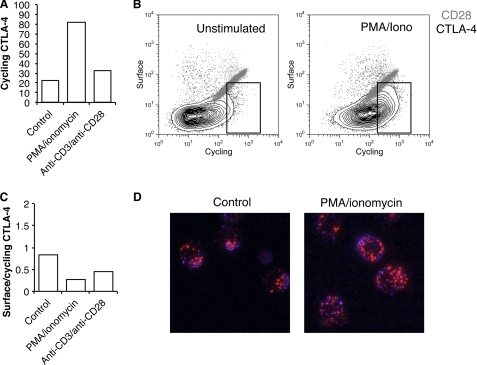
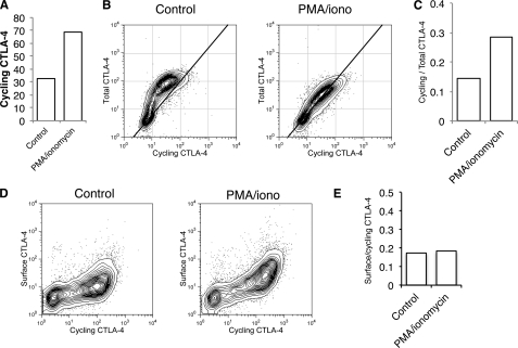
CTLA-4 is one of the most important negative regulators of the T cell immune response. However, the subcellular distribution of CTLA-4 is unusual for a receptor that interacts with cell surface transmembrane ligands in that CTLA-4 is rapidly internalized from the plasma membrane. It has been proposed that T cell activation can lead to stabilization of CTLA-4 expression at the cell surface. Here we have analyzed in detail the internalization, recycling, and degradation of CTLA-4. We demonstrate that CTLA-4 is rapidly internalized from the plasma membrane in a clathrin- and dynamin-dependent manner driven by the well characterized YVKM trafficking motif. Furthermore, we show that once internalized, CTLA-4 co-localizes with markers of recycling endosomes and is recycled to the plasma membrane. Although we observed limited co-localization of CTLA-4 with lysosomal markers, CTLA-4 was nonetheless degraded in a manner inhibited by lysosomal blockade. T cell activation stimulated mobilization of CTLA-4, as judged by an increase in cell surface expression; however, this pool of CTLA-4 continued to endocytose and was not stably retained at the cell surface. These data support a model of trafficking whereby CTLA-4 is constitutively internalized in a ligand-independent manner undergoing both recycling and degradation. Stimulation of T cells increases CTLA-4 turnover at the plasma membrane; however, CTLA-4 endocytosis continues and is not stabilized during activation of human T cells. These findings emphasize the importance of clathrin-mediated endocytosis in regulating CTLA-4 trafficking throughout T cell activation.


Figures

FIGURE 1.
FIGURE 1.
Endocytosis of CTLA-4. A, CTLA-4 C-terminal sequence alignments show the truncations used in this study. B, CHO cells expressing either wild type (wt), Δ13, or Δ23 CTLA-4 were incubated with unlabeled anti-CTLA-4 at 37 °C for 30 min. Cells were then cooled to 4 °C, and CTLA-4 that remained on the cell surface were stained with a fluorescently labeled secondary antibody (red). Cells were then fixed, permeabilized, and stained with a different secondary antibody (green) and imaged by confocal microscopy. The ratio of plasma membrane to internalized CTLA-4 fluorescence (PM/I) was calculated by outlining cells in ImageJ and is shown in the right panel. C, diagram of the antibody labeling strategy for flow cytometry experiments in D and E. D, CHO cells expressing different CTLA-4 truncations were labeled with anti-CTLA-4 PE at 37 °C for 30 min followed by labeling surface CTLA-4 on ice (4 °C) with a fluorescently conjugated anti-mouse secondary antibody. Cells were analyzed by flow cytometry. E, CHO cells expressing CD28, CTLA-4 wt, or CTLA-4 AVKM were labeled as described in C and D and analyzed by flow cytometry. Red lines indicate the staining ratio of Δ23 (D) or CD28 (E) as a guide for the expected ratio for a cell surface protein. F, CHO cells expressing wild-type CTLA-4 were labeled at 4 °C with anti-CTLA-4 PE to label surface CTLA-4. Cells were then warmed to 37 °C for the time indicated, and CTLA-4 remaining on the surface was detected on ice with a fluorescently conjugated anti-mouse secondary antibody. The time course of surface labeling is plotted against initial labeling.
FIGURE 2.
FIGURE 2.
Dynamin-dependent endocytosis of CTLA-4. A, CHO cells expressing CTLA-4 were dye-labeled (blue) and preincubated with dynasore (80 μm) for 30 min. Cells were then incubated with anti-CTLA-4 PE (red) at 37 °C in the presence of 80 μm dynasore for 30 min. B, CHO cells expressing CTLA-4 were labeled with anti-CTLA-4 PE at 37 °C for 30 min in the presence or absence of dynasore followed by labeling of surface CTLA-4 on ice with a fluorescently conjugated anti-mouse secondary antibody and analyzed by flow cytometry. C, CHO cells expressing CTLA-4 were transfected with either dynamin K44A RFP or dynamin RFP. Surface CTLA-4 was then labeled on ice using an anti-CTLA-4 APC conjugate and detected by flow cytometry. D, cells as described in C were incubated with unconjugated mouse anti-CTLA-4 for 30 min at 37 °C. Cells were then cooled to 4 °C, and receptors remaining at the cell surface were stained using an anti-mouse Alexa647 antibody (blue). Cells were fixed/permeabilized, and internalized receptors were stained with an anti-mouse Alexa555 antibody (green). Cells were analyzed by confocal microscopy.
FIGURE 3.
FIGURE 3.
Clathrin-dependent endocytosis of CTLA-4. A, CHO cells expressing CTLA-4 were transfected with GFP-tagged clathrin light chain (green). Cells were fixed and surface CTLA-4-labeled with anti-CTLA-4 Alexa546 (red). Cells were then analyzed by TIRF microscopy for co-localization of CTLA-4 and clathrin (yellow). B, CHO cells expressing CTLA-4 were transfected with either AP180C-GFP or unfused-GFP. Cells were then stained for surface CTLA-4 at 4 °C with anti-CTLA-4 PE and analyzed by flow cytometry. C, cells in B were incubated with unconjugated mouse anti-CTLA-4 for 30 min at 37 °C. Cells were then cooled to 4 °C, and receptors remaining at the cell surface were stained using an anti-mouse Alexa647 antibody (blue). Cells were then fixed and permeabilized, and internalized receptors were stained with an anti-mouse Alexa555 antibody (green). Cells were analyzed by confocal microscopy for colocalization (cyan). Right panels show enlargements of the boxed area. D, the graph shows the ratio of surface to internalized CTLA-4 fluorescence quantified from confocal images stained as in C. E, CHO cells expressing CTLA-4 (CHO CTLA-4) were transfected with RFP-tagged dominant negative inhibitors of clathrin-mediated endocytosis, and surface CTLA-4 expression was analyzed by flow cytometry by gating on RFP+ and RFP− cells from the same culture. F, CHO CTLA-4 were transfected with shRNA against the μ2 subunit of AP-2 or control shRNA constructs. Cells were labeled with anti-CTLA-4 PE at 37 °C followed by labeling surface CTLA-4 with anti-mouse 647 at 4 °C. The graph shows the ratio of surface CTLA-4 to cycling CTLA-4.
FIGURE 4.
FIGURE 4.
Internalized CTLA-4 recycles to the plasma membrane. A, CHO cells expressing CTLA-4 were transfected with Rab11-mCherry (red). Cells were then incubated with Alexa488-conjugated anti-CTLA-4 (green) for 30 min at 37 °C and observed by confocal microscopy. The white arrowheads indicate co-localization. B, CHO cells expressing CTLA-4 were labeled for 1 h with unconjugated mouse anti-CTLA-4. Cells were then placed on ice and surface CTLA-4-labeled with Alexa488 anti-mouse IgG (green). To observe recycling receptors, cells were then washed and labeled with Alexa555 anti-mouse IgG (red) at 37 °C for 45 min and fixed before confocal analysis. Control conditions are shown (left) where the Alexa555 antibody was incubated at 4 °C compared with incubation at 37 °C (right). C, flow cytometric analysis of recycling CTLA-4 is shown. Cells were labeled with mouse anti-CTLA-4 PE at 37 °C to detect cycling CTLA-4, which was followed by labeling of re-cycling protein with Alexa647 anti-mouse IgG at 4 or 37 °C for the indicated time-points. D, live-cell confocal imaging of recycling receptors was carried out using CTLA-4-expressing CHO cells. Cells were incubated with Alexa488-conjugated anti-CTLA-4 for 1 h at 37 °C. Cells were then washed, and surface CTLA-4 receptors were blocked with unconjugated anti-human IgG. Cells were then incubated with Alexa555-conjugated anti-human IgG to detect recycling receptors. Confocal Z-stacks were then acquired at the time points shown. Panel E shows quantification of cells from D. Cells were outlined in Image J, and the recycling mean pixel fluorescence is plotted against time. Error bars show S.E.
FIGURE 5.
FIGURE 5.
Degradation of CTLA-4 in lysosomal compartments. A, human primary T cell blasts were fixed and stained for CTLA-4 (green) and LAMP-1 (red) and imaged by confocal microscopy. B, HeLa cells transfected with CTLA-4 were fixed and stained for CTLA-4 (red) and anti-human LAMP-1 (green) and imaged by confocal microscopy. The right panel shows an enlarged image. C, CHO cells expressing CTLA-4 were treated as shown for 3 h followed by fixation and staining for total cellular CTLA-4 using anti-CTLA-4 PE. Cells were analyzed by flow cytometry, and the relative fluorescence was plotted. D, CHO-CTLA-4 cells were incubated in the presence of CHX or ammonium chloride (NH4Cl) for 3 h. CTLA-4 was then immunoprecipitated, and expression was analyzed by Western blotting. E, CHO-CTLA-4 cells were labeled with a 1-h pulse of anti-CTLA-4 PE. Cells were then washed and incubated for various time-points in the presence or absence of CHX and/or NH4Cl and analyzed for CTLA-4 expression by flow cytometry.
FIGURE 6.
FIGURE 6.
CTLA-4 remains predominantly intracellular in activated T cells. A, human CD4+ T cell blasts were generated using anti-CD3 anti-CD28 beads for 4 days. Cells were then incubated with anti-CD4 FITC and PE-conjugated antibody against CTLA-4 (A) or CD28 (B) at 37 °C for 60 min and visualized by confocal microscopy. C, surface CTLA-4 was detected at 4 °C. Cells were then fixed and permeabilized and stained for clathrin and analyzed by confocal microscopy.
FIGURE 7.
FIGURE 7.
Stimulation of T cell blasts leads to a mobilization but not stabilization of CTLA-4 at the cell surface. A, human CD4+ T cell blasts were generated using anti-CD3 anti-CD28 beads for 4 days. Cells were then isolated and re-stimulated with PMA+ ionomycin or fresh anti-CD3 anti-CD28 beads for 3 h at 37 °C in the presence of anti-CTLA-4 PE before analysis by flow cytometry. B, T cell blasts were re-stimulated as in A in the presence of anti-CTLA-4 PE or anti-CD28 PE at 37 °C. Cells were then placed on ice, and the remaining surface receptors were labeled by incubation with Alexa647-conjugated anti-mouse secondary antibody. Cells were analyzed by flow cytometry. The boxed area indicates an increase in CTLA-4 labeling due to stimulation. C, the ratio of surface to labeled fluorescence for cells labeled in B is plotted. D, cells stained for CTLA-4 as in B were fixed and then visualized by confocal microscopy for 37 °C staining (red) and surface staining (blue).
FIGURE 8.
FIGURE 8.
Stimulation increases trafficking of CTLA-4 in the absence of increased synthesis. A, CTLA-4-expressing Jurkat cells were labeled at 37 °C with anti-CTLA-4 PE for 1 h after stimulation with PMA/ionomycin and analyzed by flow cytometry. B, Jurkat cells were labeled at 37 °C with anti-CTLA-4 PE in the presence or absence of PMA/ionomycin for 60 min (cycling CTLA-4). Cells were then fixed, and total CTLA-4 was stained with a goat anti-CTLA-4 C-terminal antibody followed by Alexa633 anti-goat secondary and analyzed by flow cytometry. C, shown is the ratio of labeled to total CTLA-4 for cells, stained as in B. D, Jurkat cells were labeled at 37 °C with anti-CTLA-4 PE in the presence or absence of PMA/ionomycin for 60 min (cycling CTLA-4). Cells were then placed on ice, and the remaining surface receptors were labeled by incubation with Alexa647-conjugated anti-mouse secondary antibody (surface CTLA-4). Cells were analyzed by flow cytometry. E, shown is the ratio of surface to cycling CTLA-4 fluorescence for cells labeled in D.

References

    1. Tivol E. A., Borriello F., Schweitzer A. N., Lynch W. P., Bluestone J. A., Sharpe A. H. (1995) Loss of CTLA-4 leads to massive lymphoproliferation and fatal multiorgan tissue destruction, revealing a critical negative regulatory role of CTLA-4. Immunity 3, 541–547 - PubMed
    1. Waterhouse P., Penninger J. M., Timms E., Wakeham A., Shahinian A., Lee K. P., Thompson C. B., Griesser H., Mak T. W. (1995) Lymphoproliferative disorders with early lethality in mice deficient in Ctla-4. Science 270, 985–988 - PubMed
    1. Sansom D. M. (2000) CD28, CTLA-4, and their ligands. Who does what and to whom? Immunology 101, 169–177 - PMC - PubMed
    1. Teft W. A., Kirchhof M. G., Madrenas J. (2006) A molecular perspective of CTLA-4 function. Annu. Rev. Immunol. 24, 65–97 - PubMed
    1. Rudd C. E. (2008) The reverse stop-signal model for CTLA4 function. Nat. Rev. Immunol. 8, 153–160 - PubMed

Publication types

LinkOut - more resources