. 2012 Mar 16;287(12):9147-67.
doi: 10.1074/jbc.M111.316554. Epub 2012 Jan 19.

Serine/threonine phosphatase (SP-STP), secreted from Streptococcus pyogenes, is a pro-apoptotic protein

Affiliations

Serine/threonine phosphatase (SP-STP), secreted from Streptococcus pyogenes, is a pro-apoptotic protein

Shivani Agarwal et al. J Biol Chem. .

Abstract

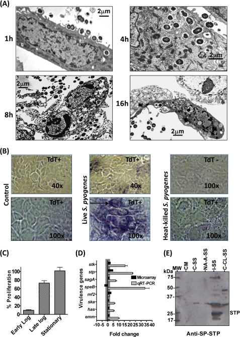
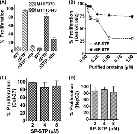
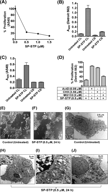
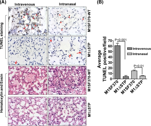
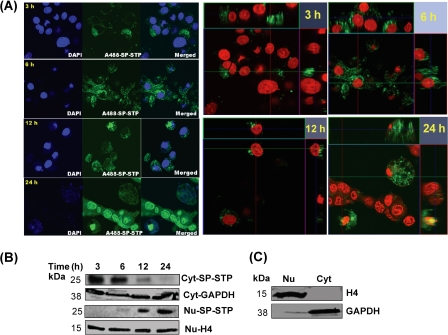
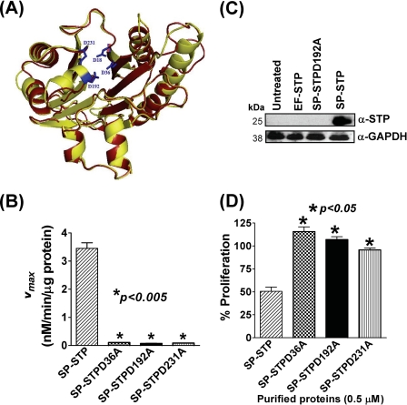
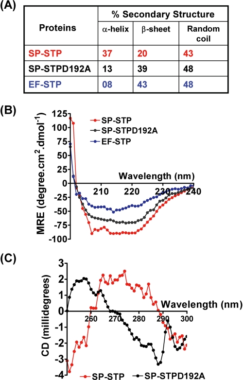
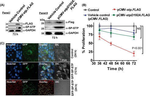
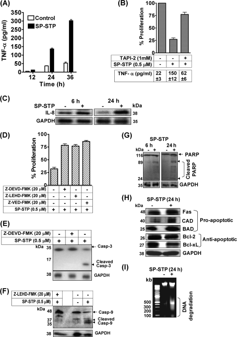
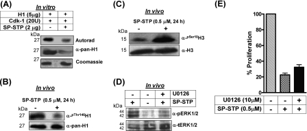
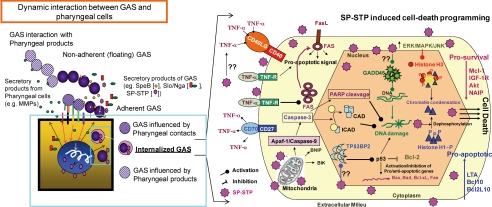
This investigation illustrates an important property of eukaryote-type serine/threonine phosphatase (SP-STP) of group A Streptococcus (GAS) in causing programmed cell death of human pharyngeal cells. The secretory nature of SP-STP, its elevated expression in the intracellular GAS, and the ability of wild-type GAS but not the GAS mutant devoid of SP-STP to cause apoptosis of the host cell both in vitro and in vivo suggest that GAS deploys SP-STP as an important virulence determinant to exploit host cell machinery for its own advantage during infection. The exogenously added SP-STP is able to enter the cytoplasm and subsequently traverses into the nucleus in a temporal fashion to cause apoptosis of the pharyngeal cells. The programmed cell death induced by SP-STP, which requires active transcription and de novo protein synthesis, is also caspase-dependent. Furthermore, the entry of SP-STP into the cytoplasm is dependent on its secondary structure as the catalytically inactive SP-STP with an altered structure is unable to internalize and cause apoptosis. The ectopically expressed wild-type SP-STP was found to be in the nucleus and conferred apoptosis of Detroit 562 pharyngeal cells. However, the catalytically inactive SP-STP was unable to cause apoptosis even when intracellularly expressed. The ability of SP-STP to activate pro-apoptotic signaling cascades both in the cytoplasm and in the nucleus resulted in mitochondrial dysfunctioning and perturbation in the phosphorylation status of histones in the nucleus. SP-STP thus not only functions as a virulence regulator but also as an important factor responsible for host-related pathogenesis.


Figures

FIGURE 1.
FIGURE 1.
Role of SP-STP in GAS-mediated apoptosis. A, transmission electron microscopy of Detroit 562 cells infected and co-cultured with mid-log phase grown M1SF370 for different incubation times as indicated. Scale bars show indicated measurement. B, TUNEL assay (DNA fragmentation) demonstrating the ability of M1SF370 GAS (live versus heat-killed) to cause apoptosis of Detroit 562 cells. C, MTT assay-based determination of percentage proliferation of Detroit 562 cells in the presence of early log, late log, and stationary phase grown M1SF370. The % proliferation inhibition was calculated by considering proliferation of untreated cells as 100%. D, comparison of the expression levels of selected virulence-related genes by microarray and qRT-PCR analyses for M1SF370 GAS population found exclusively within Detroit 562 human pharyngeal cells. E, comparison of the influence of extracellular environment and intracellular content of Detroit 562 cells on the ability of GAS to express and secrete SP-STP. Western blot analysis of the TCA-precipitated samples (Samples 1–5) obtained as described under “Experimental Procedures” and probed with anti-SP-STP antibody. The migration of molecular weight marker is shown as MW; 1st lane, CM-Tissue culture medium control; 2nd lane, C-SS-GAS in MEM without Detroit 562 cells; 3rd lane, NA-A-SS-Co-culture supernatant containing nonadherent GAS and secreted products of adherent GAS; 4th lane, I-SS-GAS incubated with cell-free extract (whole cell lysate of Detroit 562 cells); and 5th lane, C-CL-SS-control cell lysate obtained from cultured Detroit 562 cells incubated without GAS.
FIGURE 2.
FIGURE 2.
Cell type-specific role of SP-STP in mediating apoptosis. A, comparison of the ability of the wild-type (M1SF370 and M1T15448), isogenic nonpolar SP-STP-knock-out mutants (M1ΔSTP and M1T1ΔSTP), and the STP mutants complemented with the stp gene (M1ΔSTP::stp and M1T1ΔSTP::stp) (14) to inhibit proliferation of Detroit 562 cells infected at a multiplicity of infection of 100:1 (GAS CFUs:Detroit 562 cells) as revealed by MTT assay. B, MTT assay demonstrating the dose-dependent effect of the purified recombinant SP-STP and EF-STP on the proliferation of pharyngeal cells. MTT assay demonstrating the dose-dependent effect of the purified recombinant SP-STP on the proliferation of Cal-27 (C) and HepG2 cells (D).
FIGURE 3.
FIGURE 3.
SP-STP-mediates apoptosis and not necrosis of human respiratory cells. A, MTT assay demonstrating the dose-dependent effect of the purified recombinant SP-STP on the proliferation of A549. B and C, absorbance (405 nm)-based cell-death detection assay depicting the presence or absence of nucleosomal fragments in the culture supernatant (CS) and corresponding cell lysates (CL) of Detroit 562 (B) and A549 cells (C) to monitor the extent of necrosis and apoptosis in SP-STP-treated cells. D, MTT assay demonstrating the effect of transcription inhibitor actinomycin D (ActD), protein synthesis inhibitor cycloheximide (CHX), and proteasome inhibitor (MG132) on SP-STP-mediated proliferation inhibition of Detroit 562 cells. The proliferation of cells treated with the inhibitors alone in each case was taken as control. E and F, differential interference contrast images obtained by light microscopic analysis of pharyngeal cells upon SP-STP (0.5 μm, 24 h) treatment. Ultrastructural analysis of untreated (G) and SP-STP-treated (H–J) Detroit 562 cells. Apoptotic Detroit 562 cells showing: H, severely damaged mitochondria; I, nuclear indentations, prominent autophagic membranes/vacuoles (dotted circles), and incipient chromatin condensation. J, late-stage apoptotic cell with loss of cellular organelles. Scale bars show indicated measurement.
FIGURE 4.
FIGURE 4.
Biochemical basis of SP-STP-mediated apoptosis of Detroit 562 cells. A, flow cytometric analysis of the annexin-V-FITC-stained SP-STP and EF-STP treated (at indicated concentrations) pharyngeal cells. Ten thousand events were counted, and the shift in the FL-1 quadrant in comparison with the untreated cells was taken as reference to calculate percentage of apoptotic cells. B, effect of SP-STP treatment on the mitochondrial membrane potential (Ψm). Panel I, upper row shows JC-1-stained control cells showing green, red orange, and merged yellow fluorescent aggregates. The lower panel shows SP-STP-treated cells showing diffused green fluorescence dispersed throughout the cytoplasm with minimal red orange fluorescent aggregates. Panel II, quantitative assessment as measured by the ratio of red versus green (hyperpolarized versus depolarized) fluorescence in the untreated versus the SP-STP-treated (0.5 μm, 24 h) pharyngeal cells. A.U., arbitrary units. C, in vitro wound healing scratch assay showing the extent of cell migration and closing of the wound (distance in micrometers in untreated (control) and SP-STP treated (0.5 μm, 24 h) pharyngeal cells within 24 h.
FIGURE 5.
FIGURE 5.
TUNEL assay and histopathology of lungs of mice infected with wild-type (M1SF370) and isogenic mutant (M1ΔSTP) GAS strains. A, TUNEL and H&E staining of lungs of mice infected (intravenous or intranasal) with M1SF370 or M1ΔSTP (red arrows/arrowheads) show representative TUNEL-positive alveolar cells in brown. B, average TUNEL-positive cells per field obtained from three independent tissue sections.
FIGURE 6.
FIGURE 6.
A, temporal migration of AlexaFluor488-SP-STP in pharyngeal cells over a period of 24 h. The two-dimensional and the Z-stack images depict the migration of SP-STP. Nucleus is stained with Hoechst33342. The blue channel was changed to red for showing the merged fluorescence (yellow) in the Z-stacks. B, Western immunoblots showing temporal migration of SP-STP from cytoplasmic (Cyt) to nuclear (Nu) compartments as revealed by anti-SP-STP antibody. C, cytoplasmic GAPDH and nuclear histone H4 are shown as loading and purity controls.
FIGURE 7.
FIGURE 7.
A, superimposed three-dimensionally derived structures of SP-STP (yellow, active site residues highlighted in blue) and EF-STP (red) based on S. agalactiae STP (30) (Protein Data Bank code 2PK0; MMDB code 46589). B, kinetic analysis showing the catalytic coefficients (Vmax in μm/min/μg protein) of the wild-type SP-STP and its mutant proteins. C, Western blot analysis of the cell lysates harvested from SP-STP-, SP-STPD192A-, and EF-STP-treated pharyngeal cells and probed with anti-SP-STP antibody revealing the ability of the purified proteins to internalize the pharyngeal cells. Cell lysate prepared from untreated pharyngeal cells was taken as control, and GAPDH was used as loading control. Cell fractionation was carried out as in Fig. 6B. D, MTT assay showing the effect of wild-type SP-STP and the catalytically inactive mutant proteins on proliferation of Detroit 562 cells.
FIGURE 8.
FIGURE 8.
A, tabulated; B, graphical presentation of the results showing the secondary structure determination for wild-type SP-STP, SP-STPD192A, and EF-STP as determined by CD spectroscopy. MRE, mean residue ellipticity. C, tertiary structural changes in the wild-type and SP-STPD192A as determined using CD spectroscopy.
FIGURE 9.
FIGURE 9.
A, Western blot analysis using anti-FLAG and anti-SP-STP antibodies to depict the expression of STP-FLAG (Panel I) and SP-STPD192A (Panel II) 72 h post-transfection in Detroit 562 cell lysates. B, MTT assay depicting the proliferation index of the Detroit 562 cells transfected with pCMV-FLAG (vehicle control), pCMV.stp-FLAG, and pCMV-stpD192A.FLAG at the indicated time points. C, confocal microscopic differential interference contrast (DIC) and merged images showing cytoplasmic localization of GFP (24 h) and nuclear localization of SP-STP-GFP (12 and 24 h). Nucleus is stained with Hoechst33342. Blue DAPI was changed to the red channel for clarity of the superimposed images.
FIGURE 10.
FIGURE 10.
A, quantitation of TNF-α released upon SP-STP addition by ELISA. B, effect of TNF-α inhibitor, TAPI-2, on the SP-STP-induced release of TNF-α from and apoptosis of Detroit 562 cells as revealed by MTT assay. The proliferation of cells treated with the inhibitor alone was taken as control. The table below the graph depicts the amount of TNF-α released (in picograms/ml) from the untreated, SP-STP-treated, and TAPI-2-treated Detroit 562 human pharyngeal cells. C, Western blot analysis showing changes in the expression of IL-8 after 6 and 24 h of treatment. D, effect of caspase inhibitors on the SP-STP-mediated apoptosis of Detroit 562 as determined by MTT assay. E and F, SP-STP-induced cell death is caspase-dependent. Western blots depicting the SP-STP induced cleavage of caspase-3 (E) and caspase-9 (F) in the presence and absence of specific inhibitors. The proliferation of cells treated with the inhibitors alone in each case was taken as control. G, Western blot depicting the cleavage of PARP after 6 and 24 h post-SP-STP addition. H, Western blot analysis showing the changes in the expression of indicated pro- and anti-apoptotic proteins using specific antibodies as described. The GAPDH was used as loading control. I, ethidium bromide-stained 0.8% agarose gel showing DNA fragmentation patterns in SP-STP-treated (+) and untreated (−) pharyngeal cells. The first lane depicts the migration pattern of the DNA molecular weight ladder.
FIGURE 11.
FIGURE 11.
A, autoradiogram and Coomassie staining showing the ability of SP-STP to dephosphorylate the CDK-1-phosphorylated histone H1 in vitro. The identity of the samples was ascertained by immunoblotting using anti-pan-H1 antibody. B, in vivo analysis of H1 phosphorylation in the nuclear extracts isolated from the control and SP-STP-treated pharyngeal cells using anti-phospho-Thr146-H1 (α-Thr(P)-146H1) antibody. Anti-H1 antibody-reactivity (α-pan-H1) was used as the loading control. C, anti-H3-Ser(P)10 antibody (α-Ser(P)-10-H3) reactivity showing histone H3 phosphorylation in the nuclear extracts of the SP-STP-treated and -untreated (control) pharyngeal cells. Anti-H3 antibody reactivity (Nu-H3) was used as the loading control. D, Western blot analysis depicting the change in the phospho-ERK1/2 (pERK1/2) profile in the untreated and SP-STP-treated pharyngeal cells alone or in the presence of the ERK1/2 inhibitor, U0126. The level of total ERK1/2 (tERK1/2) was taken as the loading control. E, MTT assay showing the SP-STP induced-proliferation inhibition of Detroit 562 cells in the presence and absence of ERK1/2 inhibitor U0126. The proliferation of cells treated with the inhibitor alone was taken as control.
FIGURE 12.
FIGURE 12.
Model depicting the up-/down-regulation of pro-/anti-apoptotic cascades in both the cytoplasmic and nuclear compartments of the Detroit 562 human pharyngeal cells upon SP-STP treatment. GAS responds to the external environment. Hence the genes expression repertoire in GAS varies according to the constantly changing environments within the host. As a result, the gene expression profiles of the GAS population not in contact with the host cells (floating/non adherent GAS) versus the GAS population in contact with the host cells (adherent GAS) versus those that are found within the host cells (invaded GAS) are quite different. We found that the GAS found exclusively within the host cells is highly virulent as many virulence and virulence-regulating genes are up-regulated. Among them, eukaryote-like Ser/Thr phosphatase (SP-STP) is the focus of this study, which has been reported previously as a secretory protein. SP-STP, by virtue of its eukaryote-type nature and its overexpression within the cytoplasm of pharyngeal cells upon GAS invasion, becomes a promiscuous protein exploited by GAS to hijack host signal transduction cascades both extrinsically, i.e. when it is secreted from the adherent GAS, and intrinsically, when it is secreted in large amounts by the intracellular invaded GAS. We demonstrate that the final destination of SP-STP whether from outside or inside is the host cell nucleus. The Ser/thr phosphatase activity of SP-STP directly or indirectly activates expression of several pro-apoptotic genes and suppresses anti-apoptotic genes. The cumulative effects of these processes culminate in mitochondrial malfunctioning, changes in histone phosphorylation status, chromatin fragmentation, and condensation. Ultimately these changes result in apoptosis and proliferation inhibition of pharyngeal cells. SP-STP thus significantly contributes to the GAS-mediated apoptosis.

References

    1. Carapetis J. R., Steer A. C., Mulholland E. K., Weber M. (2005) The global burden of group A streptococcal diseases. Lancet Infect. Dis. 5, 685–694 - PubMed
    1. Olsen R. J., Shelburne S. A., Musser J. M. (2009) Molecular mechanisms underlying group A streptococcal pathogenesis. Cell. Microbiol. 11, 1–12 - PubMed
    1. Virtaneva K., Porcella S. F., Graham M. R., Ireland R. M., Johnson C. A., Ricklefs S. M., Babar I., Parkins L. D., Romero R. A., Corn G. J., Gardner D. J., Bailey J. R., Parnell M. J., Musser J. M. (2005) Longitudinal analysis of the group A Streptococcus transcriptome in experimental pharyngitis in cynomolgus macaques. Proc. Natl. Acad. Sci. U.S.A. 102, 9014–9019 - PMC - PubMed
    1. Goldmann O., Rohde M., Chhatwal G. S., Medina E. (2004) Role of macrophages in host resistance to group A streptococci. Infect. Immun. 72, 2956–2963 - PMC - PubMed
    1. Medina E., Rohde M., Chhatwal G. S. (2003) Intracellular survival of Streptococcus pyogenes in polymorphonuclear cells results in increased bacterial virulence. Infect. Immun. 71, 5376–5380 - PMC - PubMed

Publication types

MeSH terms

Substances