. 2012 Mar 23;287(13):10613-10622.
doi: 10.1074/jbc.M111.323501. Epub 2012 Jan 18.

Three-dimensional structure of steroid 21-hydroxylase (cytochrome P450 21A2) with two substrates reveals locations of disease-associated variants

Affiliations

Three-dimensional structure of steroid 21-hydroxylase (cytochrome P450 21A2) with two substrates reveals locations of disease-associated variants

Bin Zhao et al. J Biol Chem. .

Abstract

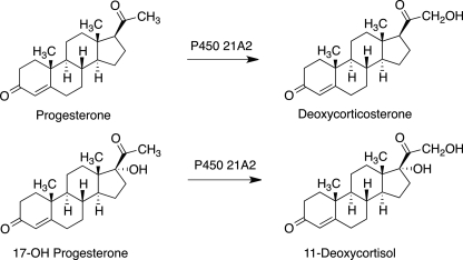
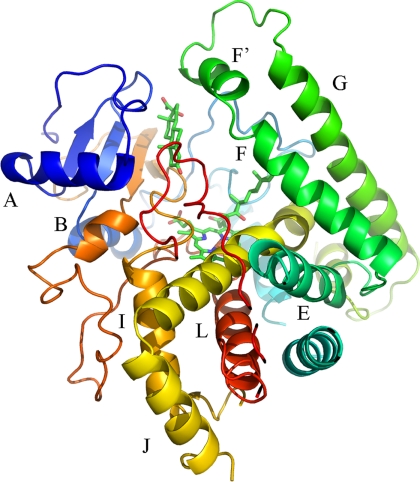
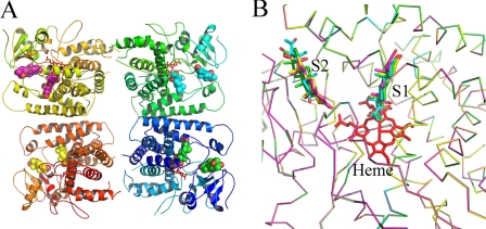
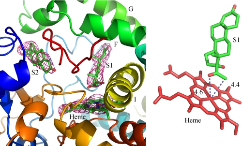
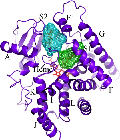
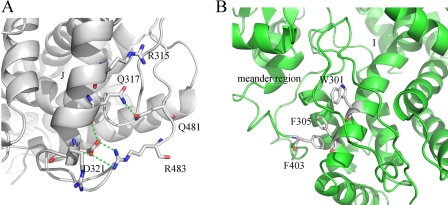
Steroid 21-hydroxylase (cytochrome P450 21A2, CYP21A2) deficiency accounts for ∼95% of individuals with congenital adrenal hyperplasia, a common autosomal recessive metabolic disorder of adrenal steroidogenesis. The effects of amino acid mutations on CYP21A2 activity lead to impairment of the synthesis of cortisol and aldosterone and the excessive production of androgens. In order to understand the structural and molecular basis of this group of diseases, the bovine CYP21A2 crystal structure complexed with the substrate 17-hydroxyprogesterone (17OHP) was determined to 3.0 Å resolution. An intriguing result from this structure is that there are two molecules of 17OHP bound to the enzyme, the distal one being located at the entrance of the substrate access channel and the proximal one bound in the active site. The substrate binding features locate the key substrate recognition residues not only around the heme but also along the substrate access channel. In addition, orientation of the skeleton of the proximal molecule is toward the interior of the enzyme away from the substrate access channel. The 17OHP complex of CYP21A2 provides a good relationship between the crystal structure, clinical data, and genetic mutants documented in the literature, thereby enhancing our understanding of congenital adrenal hyperplasia. In addition, the location of certain CYP21A2 mutations provides general understanding of structure/function relationships in P450s.


Figures

FIGURE 1.
FIGURE 1.
Reactions catalyzed by CYP21A2.
FIGURE 2.
FIGURE 2.
Ribbon diagram of the 17OHP-bound engineered CYP21A2 (C3B21RA) structure. The heme and the two 17OHP molecules are colored green in stick models. The F helix is stretched into two short helices, and the F′/G helices swing away from the structural core.
FIGURE 3.
FIGURE 3.
Evidence for the presence of four molecules present in the asymmetric unit cell. A, the four CYP21A2 structures are colored in green, yellow, blue, and orange. The two molecules of 17OHP are seen in cyan, pink, green, and yellow as a sphere model. Heme is in a red stick model. B, superimposed overall structures of 17OHP-bound CYP21A2 as a backbone stick model. All four proximal 17OHP molecules are in the same orientation, and the locations of the four distal 17OHP molecules show small variations in binding modes in the substrate access channel.
FIGURE 4.
FIGURE 4.
Electron density for 17OHP in the structure of 17OHP-bound CYP21A2. The electron density map was calculated using σA-weighted 2|Fo| − |Fc| coefficients and is contoured at 1.0 σ. The proximal 17OHP and distal 17OHP molecules in the green stick models (left) are denoted S1 and S2, respectively. Potential hydrogen bonds and the distances between the corresponding carbon and oxygen atoms to the iron atom are shown as dotted lines (right).
FIGURE 5.
FIGURE 5.
Solvent-accessible surfaces of the substrate binding cavities of CYP21A2. The cavities are shown as mesh surfaces. The distal binding site in the substrate access channel is in cyan, and the proximal binding site in the heme pocket is green. These two binding sites are connected by a narrow channel.
FIGURE 6.
FIGURE 6.
Steady-state binding of 17OHP to CYP21A2. Two 1.0-ml cuvettes each contained 2.0 μm P450 21A2 in 50 mm potassium phosphate buffer (pH 7.4). A base line was recorded (OLIS-Amico DW2a spectrophotometer), and 1-μl aliquots of 400 μm 17OHP (in C2H5OH) were added to the sample cuvette, with an equivalent amount of C2H5OH added to the reference cuvette at each step (B, inset). The points (ΔA390A418) were fit (using the program DynaFit) to a two-site model with Kd,1 = 0.05 μm, Kd,2 = 0.7 μm (A) (using Δϵ1 = Δϵ2 = 26 mm−1 cm−1) (supplemental Fig. S1). See Fig. 7 for pre-steady-state data of spectral changes.
FIGURE 7.
FIGURE 7.
Pre-steady-state binding of progesterone and 17OHP to CYP21A2. Spectra were recorded in an OLIS-RSM 1000 instrument at 37 °C. One syringe contained 4 μm P450 21A2 in 50 mm potassium phosphate buffer (pH 7.4), 100 mm NaCl, 0.1 mm dithiothreitol, and 0.1 mm EDTA. The other syringe contained 40 μm progesterone or 17OHP in the same buffer. The components (equal volumes) were mixed, and spectra were acquired every 1 ms. Some of the early traces and the final trace (16 s) are shown in A (fully changed). ΔA390 data points were fit to biexponential plots using the manufacturer's software (GlobalWorks). B, progesterone binding. k1 = 1.8 ± 0.7 s−1, k2 = 0.062 ± 0.06 s−1. C, 17OHP binding. k1 = 2.5 ± 0.3 s−1, k2 = 0.26 ± 0.10 s−1.
FIGURE 8.
FIGURE 8.
Detailed local structural features of CYP21A2. A, Gly-65 (in a van der Waals surface model) is located at the β-hairpin turn and is less than 4 Å from the distal substrate. It serves as a gating residue at the mouth of the substrate access channel; B, Glu-379 is directed toward the surface of the protein and at the loop between β1-3 and β1-4. The carbonyl group of Glu-379 can form a hydrogen bond with the oxygen of Thr-367, thus locking the two β sheets in the structure. Thr-367 makes an additional hydrogen bond with Asp-88, which is in the loop near the B helix. These hydrogen bond interactions play an important role in maintaining the structural fold. Dotted lines show the presumed hydrogen bonding network.
FIGURE 9.
FIGURE 9.
The continuous hydrogen bonding network in the engineered CYP21A2 complexes. A, Arg-483 and Gln-481 lie near the C-terminal end of the protein. The side chains of both Arg-483 and Gln-481 form hydrogen bonds with Asp-321 and Gln-317 in the J helix; B, the side chains of Trp-301, Phe-305, and Phe-403 form π-electron stacking interactions, thus linking the meander region and the C terminus of the I helix and stabilizing the structure.

References

    1. Kagawa N., Waterman M. R. (1991) Evidence that an adrenal-specific nuclear protein regulates the cAMP responsiveness of the human CYP21B (P450C21) gene. J. Biol. Chem. 266, 11199–11204 - PubMed
    1. Kagawa N., Waterman M. R. (1990) cAMP-dependent transcription of the human CYP21B (P-450C21) gene requires a cis-regulatory element distinct from the consensus cAMP-regulatory element. J. Biol. Chem. 265, 11299–11305 - PubMed
    1. White P. C., Speiser P. W. (2000) Congenital adrenal hyperplasia due to 21-hydroxylase deficiency. Endocr. Rev. 21, 245–291 - PubMed
    1. Speiser P. W., White P. C. (2003) Congenital adrenal hyperplasia. N. Engl. J. Med. 349, 776–788 - PubMed
    1. White P. C., New M. I., Dupont B. (1986) Structure of human steroid 21-hydroxylase genes. Proc. Natl. Acad. Sci. U.S.A. 83, 5111–5115 - PMC - PubMed

Publication types

MeSH terms

Substances

Associated data