. 2012 Jan 18;32(3):953-65.
doi: 10.1523/JNEUROSCI.4640-11.2012.

Cervicolumbar coordination in mammalian quadrupedal locomotion: role of spinal thoracic circuitry and limb sensory inputs

Affiliations

Cervicolumbar coordination in mammalian quadrupedal locomotion: role of spinal thoracic circuitry and limb sensory inputs

Laurent Juvin et al. J Neurosci. .

Abstract

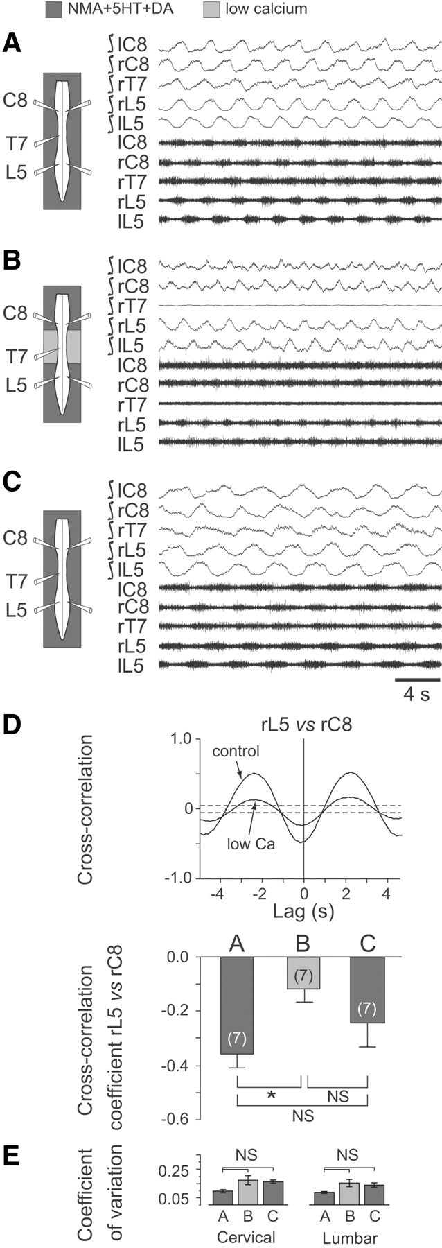
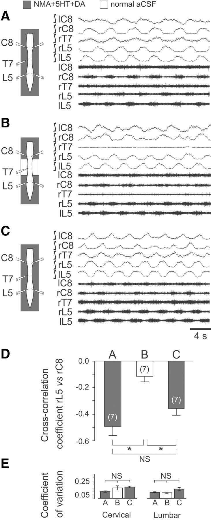
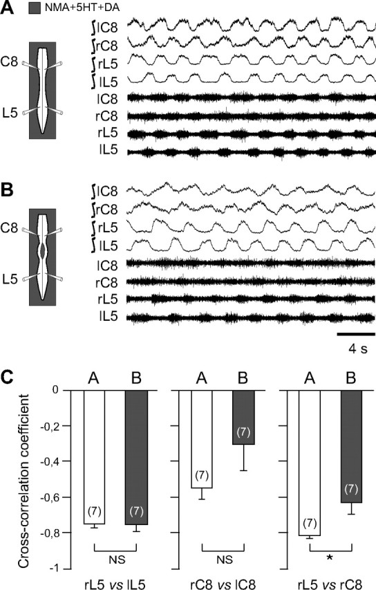
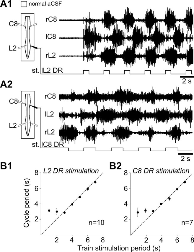
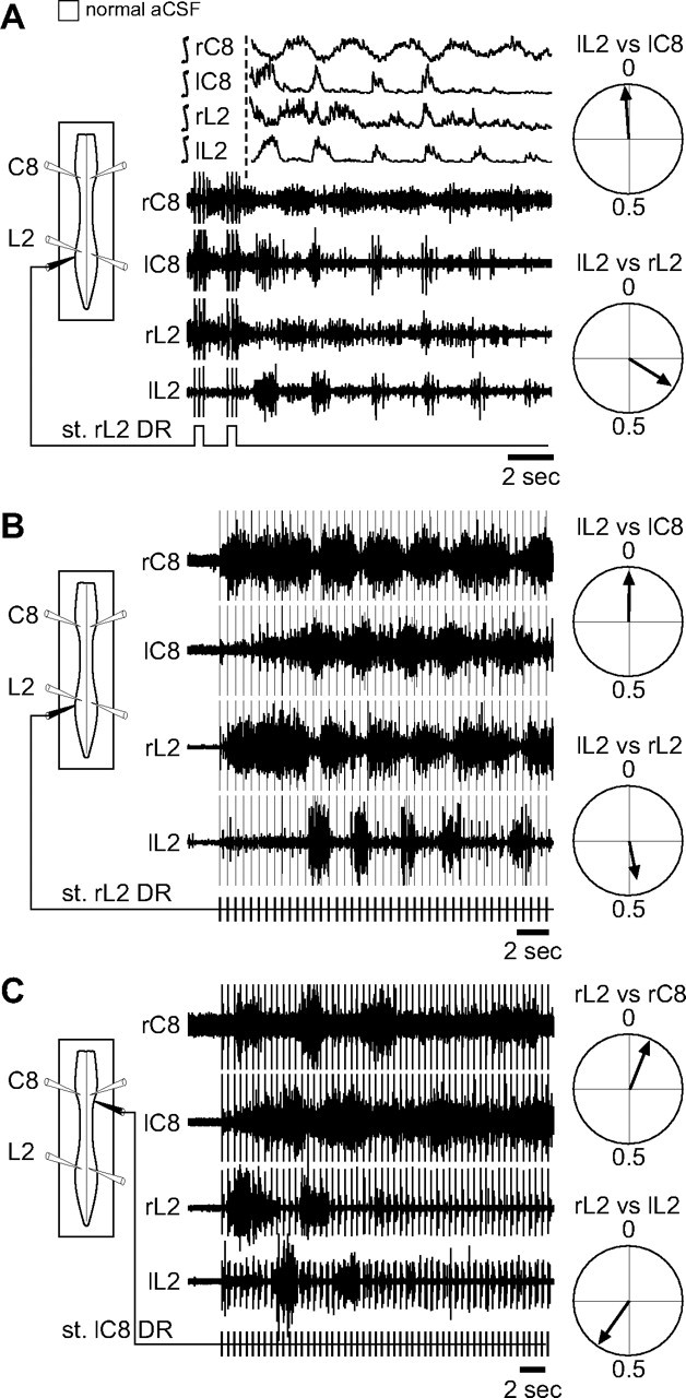
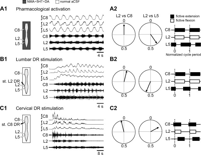
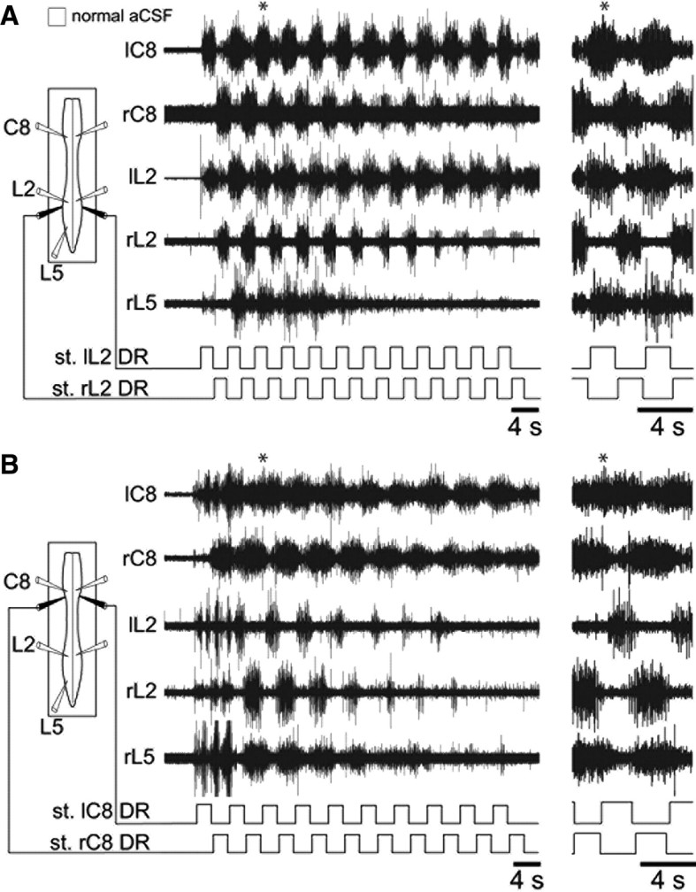
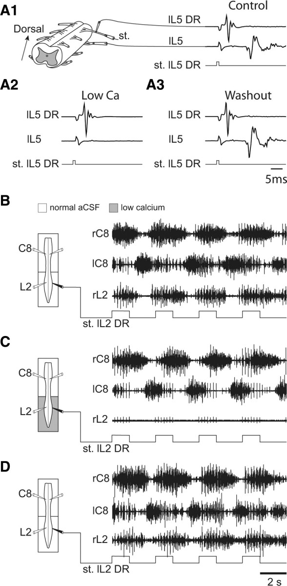
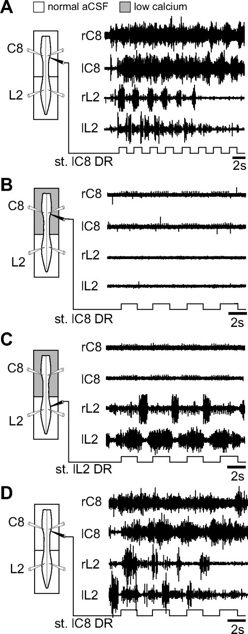
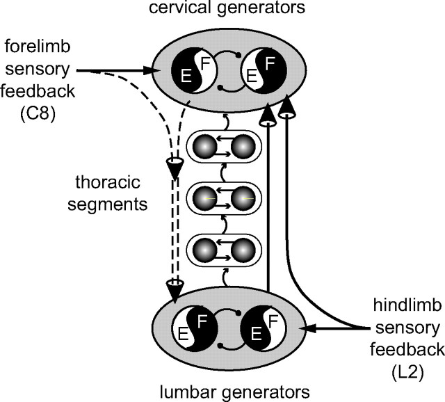
Effective quadrupedal locomotion requires a close coordination between the spatially distant central pattern generators (CPGs) controlling forelimb and hindlimb movements. Using isolated preparations of the neonatal rat spinal cord, we explore the role of intervening thoracic circuitry in cervicolumbar CPG coordination and the contribution to this remote coupling of limb somatosensory inputs. In preparations activated with bath-applied N-methyl-D,L-aspartate, serotonin, and dopamine, the coordination between locomotor-related bursts recorded in cervical and lumbar ventral roots was substantially weakened, although not abolished, when the thoracic segments were selectively withheld from neurochemical stimulation or were exposed to a low Ca(2+) solution to block synaptic transmission. Moreover, cervicolumbar CPG coordination was reduced after a thoracic midsagittal section, suggesting that cross-cord projections participate in the anteroposterior coupling. In quiescent preparations, either cyclic or tonic electrical stimulation of low-threshold afferent pathways in C8 or L2 dorsal roots (DRs) could elicit coordinated ventral root bursting at both cervical and lumbar levels via an activation of the underlying CPG networks. When lumbar rhythmogenesis was prevented by local synaptic transmission blockade, L2 DR stimulation could still drive left-right alternating cervical bursting in preparations otherwise exposed to normal bathing medium. In contrast, when the cervical generators were selectively blocked, C8 DR stimulation was unable to activate the lumbar CPGs. Thus, in the newborn rat, anteroposterior limb coordination relies on active burst generation within midcord thoracic circuitry that additionally conveys ascending and weaker descending coupling influences of distant limb proprioceptive inputs to the cervical and lumbar generators, respectively.


Figures

Figure 1.
Figure 1.
Blockade of synaptic transmission in thoracic segments of the neonatal rat spinal cord weakens the coordination between cervical (forelimb) and lumbar (hindlimb) locomotor-related rhythmicity. A–C, Left, Schematics of the isolated spinal cord with locations of extracellular electrode recordings. Right, Raw extracellular (bottom traces) and corresponding integrated activity (top traces) in left (l) and right (r) cervical (C), thoracic (T) and lumbar (L) ventral roots during fictive locomotion induced by NMA/5-HT/DA applied to the whole cord (A), during selective blockade of synaptic transmission in the thoracic cord region (from T3 to T10) with a low Ca2+/high Mg2+ medium (B), and after restoration of generalized normal ACSF conditions (C). D, Top, Cross-correlograms showing alternation (indicated by a negative cross-correlation coefficient at lag 0) between homolateral lumbar (L5) and cervical (C8) ventral root bursts in control conditions and during exposure of the thoracic segments to low Ca2+/high Mg2+ (corresponding to A and B, respectively). Note that the strength of coordination between L5/C8 motor bursts was reduced under thoracic cord synaptic blockade. Each cross-correlogram was computed from 2–3 min of activity; dashed lines indicate the 95% (±2 SEM) confidence interval. Bottom, Histograms showing the mean cross-correlation coefficients (±SEM) of homolateral L5 versus C8 ventral root bursts in the three experimental conditions illustrated in A–C. Numbers of preparations are indicated in parentheses. E, Histograms showing mean coefficients of variation (vertical bars) and corresponding SDs (vertical lines) of cervical (left) and lumbar rhythms (right) before, during, and after application of low Ca2+/high Mg2+ ACSF to the thoracic cord region. *p < 0.05. NS, Not significantly different.
Figure 2.
Figure 2.
Neurochemical activation of the thoracic cord region significantly enhances the coordination of cervicolumbar locomotor rhythmicity. A–C, Left, Schematics of experimental procedure. Right, Integrated and corresponding raw extracellular recordings from bilateral cervical (C), thoracic (T), and lumbar (L) ventral roots during bath application of NMA/5-HT/DA to the whole cord (A), to the cervical and the lumbar cord enlargements only (B), and again to the entire cord (C). In B, thoracic segments from T3 to T10 were exposed to normal ACSF. D, Mean cross-correlation coefficients (±SEM) of right lumbar (rL5) versus right cervical (rC8) ventral root activity during NMA/5-HT/DA application to the whole cord (A, C) or to cervical and lumbar spinal segments with the thoracic cord under normal ACSF (B). E, Histograms of the mean coefficients of variation (vertical bars) and corresponding SDs (vertical lines) of cervical (left) and lumbar rhythms (right) in the three experimental conditions. Numbers of preparations are indicated in parentheses. *p < 0.05. NS, Not significantly different.
Figure 3.
Figure 3.
Thoracic cross-cord connections contribute to the coordination of cervicolumbar locomotor rhythmicity. A, B, Left, Schematics of experimental procedure. Right, Integrated and corresponding raw extracellular recordings from left (l) and right (r) cervical (C8) and lumbar (L5) ventral roots during NMA/5HT/DA application to the whole cord (A) and after a thoracic midsagittal section extending from segments T3 to T10 (B). C, Mean cross-correlation coefficients (±SEM) of right versus left L5 (left histogram pair), bilateral C8 (middle histogram pair), and homolateral L5 versus C8 (right histogram pair) ventral root bursts before (unfilled bars) and after (shaded bars) a thoracic midline section as in A and B, respectively. Note that the strength of cervicolumbar burst coordination, but not of bilateral segmental coupling, was significantly affected by the interruption of cross-cord thoracic pathways. Numbers of preparations are indicated in parentheses. *p < 0.05. NS, Not significantly different.
Figure 4.
Figure 4.
Cyclic activation of low-threshold lumbar or cervical DR afferent fibers elicits coordinated cervicolumbar locomotor bursting. A1, A2, Left, Schematics indicating the positions of ventral root recording (unfilled) and DR stimulating (filled) electrodes. Right, Raw ventral root activity at cervical (C8) and lumbar (L2) levels in response to electrical train stimulation (st.) of lumbar (L2; A1) or cervical (C8; A2) DRs. B1, B2, Scatter plots of the relationship between the cycle period of rhythmic motor bursts and the period of lumbar (B1) or cervical (B2) DR stimulus trains. Each point represents the mean motor burst period during 5–10 consecutive cycles of DR stimulation. The dashed lines in B1 and B2 indicate a one-to-one coupling, which in both cases fails with stimulus periods below ∼3 s.
Figure 5.
Figure 5.
The DR stimulation-elicited motor patterns result from the activation of locomotor rhythm-generating circuitry. A, Left, Schematic of the preparation with recording electrodes (unfilled) placed on bilateral cervical (C8) and lumbar (L2) ventral roots and a stimulating electrode (filled) on right L2 DR. Middle, Integrated and raw motor activity immediately after the termination of an episode of cyclic L2 DR stimulation (st.). Note the occurrence of several cycles of locomotor-like bursting after termination of afferent input stimulation. Right, Circular plots (from 15 poststimulus cycles in 3 preparations) of the phase relationships between motor burst onsets in ipsilateral left L2 versus C8 (top) and left L2 vs right L2 (bottom) ventral roots. B, C, Left, Schematic of the experimental procedure, which was the same as in A except that the right L2 (B) or the left C8 (C) DR was now stimulated tonically (bottom traces). In both cases, well-coordinated cervicolumbar ventral root bursting was expressed in a pattern that was unrelated to the timing of DR stimulation. Right, Corresponding phase plots of L2 versus C8 (top) and left versus right L2 (bottom) ventral root bursts in the two tonic stimulus conditions. The plots in B and C were each constructed from 15 cycles expressed during stimulation-induced bursting in five and three different preparations, respectively.
Figure 6.
Figure 6.
Comparison of fictive locomotor patterns evoked either by neuroactive drug application or by electrical stimulation (st.) of low-threshold limb sensory pathways. A1, B1, C1, Left, Schematics of experimental procedure. Recording electrodes (unfilled) were placed on ipsilateral cervical (C8) and lumbar (L2, L5) ventral roots. Right, Typical coordinated motor burst patterns induced by NMA/5-HT/DA application to the whole cord (A1) or by rhythmic electrical train stimulation (filled electrode) of ipsilateral lumbar (L2; B1) or cervical (C8; C1) DRs under normal ACSF exposure. A2, B2, C2, Left, Corresponding circular plots of the phase relationships between the onsets of motor bursts recorded over ≥15 random cycles from L2 and C8 (left plots) or L2 and L5 (right plots) ventral roots in the three experimental conditions. Right, Phase diagrams showing duty cycles of ipsilateral C8, L2, and L5 ventral root bursts during normalized cycles of fictive locomotion in each case.
Figure 7.
Figure 7.
Coordinated cervicolumbar ventral root burst activation by alternating bilateral stimulation (st.) of low-threshold lumbar or cervical dorsal root afferents. A, B, Left, Schematics of experimental procedure. Middle, Raw motor activity at lumbar (left and right L2, L5) and cervical (left and right C8) levels during alternating cyclic stimulation (monitored in lower traces) of left and right L2 (A) or C8 (B) DRs. In each case, an expanded portion (indicated by asterisks) is placed at right to show greater detail. Note the occurrence of alternating (A) or overlapping (B) L2 and L5 motor bursts during the lumbar or cervical DR stimulation, respectively.
Figure 8.
Figure 8.
Direct ascending influence of low-threshold lumbar sensory afferents on the cervical pattern generators. A1–A3, Ability of low-calcium ACSF to reversibly block synaptic transmission in segmental reflex pathways. A1, A low-threshold afferent volley (top trace) recorded proximally from an L5 DR in response to a distally applied stimulus (st.) pulse (0.6 V; bottom trace) and the synaptically driven response in the L5 ventral root (middle trace). In the presence of low Ca2+/high Mg2+ (A2), the motor response to the same DR activation was abolished, but reappeared after washout with normal ACSF (A3). B–D, Left, Schematics of the isolated spinal cord with locations of recording electrodes (unfilled) placed on cervical (left and right C8) and lumbar (right L2) ventral roots, and a stimulating electrode (filled) placed on the left L2 DR. A Vaseline partition allowed the differential perfusion of the cervical and lumbar cord regions. Right, Effects of cyclic L2 DR activation on bilateral C8 and right L2 ventral root activity under control conditions (B), during exposure of the lumbar cord to low Ca2+/high Mg2+ (C), and after washout with normal ACSF (D). Robust bilaterally alternating cervical bursting persisted in response to L2 DR stimulation under the two perfusion conditions and notably in the absence of lumbar CPG activity (as in C).
Figure 9.
Figure 9.
The descending influence of low-threshold cervical afferent inputs on the lumbar locomotor CPGs requires cervical pattern generator activation. A–D, Left, Schematics of isolated preparations showing recording electrode locations (left/right C8 and L2 ventral roots) and a stimulating electrode placed on either the left C8 (A, B, D) or left L2 DRs (C). A Vaseline partition allowed the differential perfusion of the cervical and lumbar cord regions. Right, Effects of cyclic stimulation (st.) of low-threshold cervical afferents on bilateral C8 and L2 motor root activity under control conditions (A), during blocked of synaptic transmission in the cervical cord region (B), and after washout with normal ACSF (D). Note that during low Ca2+/high Mg2+ application to the anterior cord, cyclic stimulation of an L2 DR was still able to activate bilaterally alternating bursting in L2 motor roots (C).
Figure 10.
Figure 10.
Summary diagram of asymmetrical spinal pathways contributing to the coordination of the spatially distant rhythmogenic networks underlying quadrupedal locomotion in neonatal rat. In addition to direct ascending inter-network connections from the hindlimb-to-forelimb pattern generators (solid vertical line at right), anteroposterior limb coordination requires the active participation of relay circuitry in intervening thoracic cord segments (shaded circles and intersegmental arrows). Moreover, unlike the ascending influence of hindlimb sensory afferents on the cervical pattern generators, which bypasses the lumbar CPGs (second vertical line at right), the rostrocaudal impact of forelimb sensory inputs on the hindlimb generators requires the concomitant activation of the cervical CPGs (dashed vertical lines at left). See Discussion for further explanation. E, Extensor; F, flexor.

Similar articles

Cited by

References

    1. Baldissera F, Cavallari P, Leocani L. Cyclic modulation of the H-reflex in a wrist flexor during rhythmic flexion-extension movements of the ipsilateral foot. Exp Brain Res. 1998;118:427–430. - PubMed
    1. Ballion B, Morin D, Viala D. Forelimb locomotor generators and quadrupedal locomotion in the neonatal rat. Eur J Neurosci. 2001;14:1727–1738. - PubMed
    1. Bareyre FM, Kerschensteiner M, Raineteau O, Mettenleiter TC, Weinmann O, Schwab ME. The injured spinal cord spontaneously forms a new intraspinal circuit in adult rats. Nat Neurosci. 2004;7:269–277. - PubMed
    1. Barriere G, Mellen N, Cazalets JR. Neuromodulation of the locomotor network by dopamine in the isolated spinal cord of newborn rat. Eur J Neurosci. 2004;19:1325–1335. - PubMed
    1. Cazalets JR. Metachronal propagation of motoneurone burst activation in isolated spinal cord of newborn rat. J Physiol. 2005;568:583–597. - PMC - PubMed

Publication types

MeSH terms

LinkOut - more resources