

Plastin family of actin-bundling proteins: its functions in leukocytes, neurons, intestines, and cancer
- PMID: 22262972
- PMCID: PMC3259490
- DOI: 10.1155/2012/213492
Plastin family of actin-bundling proteins: its functions in leukocytes, neurons, intestines, and cancer
Abstract
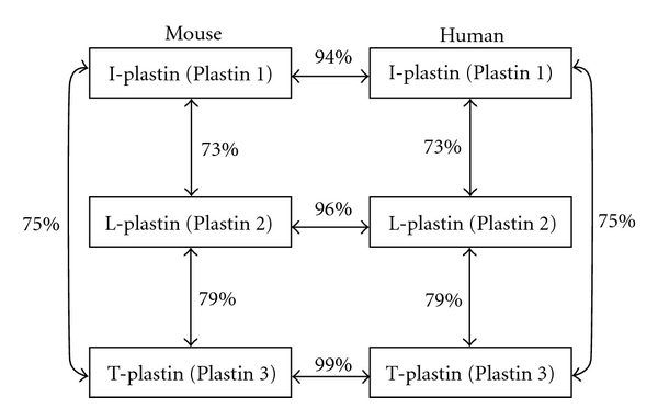
Sophisticated regulation of the actin cytoskeleton by a variety of actin-binding proteins is essential for eukaryotic cells to perform their diverse functions. The plastin (also know, as fimbrin) protein family belongs to actin-bundling proteins, and the protein family is evolutionarily conserved and expressed in yeast, plant, and animal cells. Plastins are characterized by EF-hand Ca(2+)-binding domains and actin-binding domains and can cross-link actin filaments into higher-order assemblies like bundles. Three isoforms have been identified in mammals. T-plastin is expressed in cells from solid tissues, such as neurons in the brain. I-plastin expression is restricted to intestine and kidney; the isoform plays a vital role in the function of absorptive epithelia in these organs. L-plastin is expressed in hematopoietic cell lineages and in many types of cancer cells; the isoform is thus considered to be a useful biomarker for cancer.
Figures
References
-
- Bañuelos S, Saraste M, Carugo KD. Structural comparisons of calponin homology domains: implications for actin binding. Structure. 1998;6(11):1419–1431. - PubMed
-
- Namba Y, Ito M, Zu Y, Shigesada K, Maruyama K. Human T cell L-plastin bundles actin filaments in a calcium dependent manner. Journal of Biochemistry. 1992;112(4):503–507. - PubMed
-
- Goldsmith SC, Pokala N, Shen W, Fedorov AA, Matsudaira P, Almo SC. The structure of an actin-crosslinking domain from human fimbrin. Nature Structural Biology. 1997;4(9):708–712. - PubMed
LinkOut - more resources
Full Text Sources
Other Literature Sources
Miscellaneous
