Application of Three-dimensional Reconstruction in Esophageal Foreign Bodies
- PMID: 22263191
- PMCID: PMC3249343
- DOI: 10.5090/kjtcs.2011.44.5.368
Application of Three-dimensional Reconstruction in Esophageal Foreign Bodies
Abstract
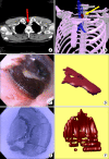
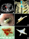
This study was conducted to investigate the clinical application of three-dimensional (3D) reconstructed computed tomography (CT) images in detecting and gaining information on esophageal foreign bodies (FBs). Two patients with esophageal FBs were enrolled for analysis. In both cases, 3D reconstructed images were compared with the FB that was removed according to the object shape, size, location, and orientation in the esophagus. The results indicate the usefulness of conversion of CT data to 3D images to help in diagnosis and treatment. Use of 3D images prior to treatment allows for rapid prototyping and surgery simulation.
Keywords: Computed tomography; Esophageal foreign body; Esophageal surgery.
Figures


Similar articles
-
Value of 3-dimensional CT virtual anatomy imaging in complex foreign body retrieval from soft tissues.Korean J Radiol. 2013 Mar-Apr;14(2):269-77. doi: 10.3348/kjr.2013.14.2.269. Epub 2013 Feb 22. Korean J Radiol. 2013. PMID: 23483807 Free PMC article.
-
Usefulness of Ultralow-Dose (Submillisievert) Chest CT Using Iterative Reconstruction for Initial Evaluation of Sharp Fish Bone Esophageal Foreign Body.AJR Am J Roentgenol. 2015 Nov;205(5):985-90. doi: 10.2214/AJR.15.14353. AJR Am J Roentgenol. 2015. PMID: 26496545
-
Management of foreign bodies in the airway and oesophagus.Int J Pediatr Otorhinolaryngol. 2012 May 14;76 Suppl 1:S84-91. doi: 10.1016/j.ijporl.2012.02.010. Epub 2012 Feb 24. Int J Pediatr Otorhinolaryngol. 2012. PMID: 22365376 Review.
-
Endoscopic management of gastrointestinal foreign bodies in children.Indian J Pediatr. 1999;66(1 Suppl):S75-80. Indian J Pediatr. 1999. PMID: 11132474 Review.
Cited by
-
The current and possible future role of 3D modelling within oesophagogastric surgery: a scoping review.Surg Endosc. 2022 Aug;36(8):5907-5920. doi: 10.1007/s00464-022-09176-z. Epub 2022 Mar 11. Surg Endosc. 2022. PMID: 35277766 Free PMC article.
-
Study of foreign-body extraction from the upper third of the esophagus in children.Iran J Pediatr. 2014 Apr;24(2):214-8. Iran J Pediatr. 2014. PMID: 25535542 Free PMC article.
-
Three-Dimensional Computed Tomography for the Safe Management of Esophageal Foreign Body.Cureus. 2025 Jul 11;17(7):e87720. doi: 10.7759/cureus.87720. eCollection 2025 Jul. Cureus. 2025. PMID: 40786442 Free PMC article.
References
-
- Young CA, Menias CO, Bhalla S, Prasad SR. CT features of esophageal emergencies. Radiographics. 2008;28:1541–1553. - PubMed
-
- Hirasaki S, Inoue A, Kubo M, Oshiro H. Esophageal large fish bone (Sea Bream Jawbone) impaction successfully managed with endoscopy and safely excreted through the intestinal tract. Intern Med. 2010;49:995–999. - PubMed
-
- Cheng HT, Wu CI, Tseng CS, et al. The occlusion-adjusted prefabricated 3D mirror image templates by computer simulation: the image-guided navigation system application in difficult cases of head and neck reconstruction. Ann Plast Surg. 2009;63:517–521. - PubMed
-
- Sannomiya EK, Silva JV, Brito AA, Saez DM, Angelieri F, Dalben Gda S. Surgical planning for resection of an ameloblastoma and reconstruction of the mandible using a selective laser sintering 3D biomodel. Oral Surg Oral Med Oral Pathol Oral Radiol Endod. 2008;106:e36–e40. - PubMed
Publication types
LinkOut - more resources
Full Text Sources

