Application of Three-dimensional Reconstruction in Esophageal Foreign Bodies
- PMID: 22263191
- PMCID: PMC3249343
- DOI: 10.5090/kjtcs.2011.44.5.368
Application of Three-dimensional Reconstruction in Esophageal Foreign Bodies
Abstract
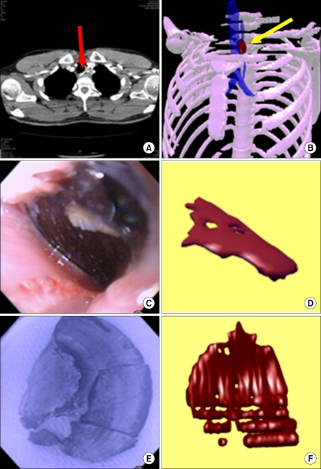
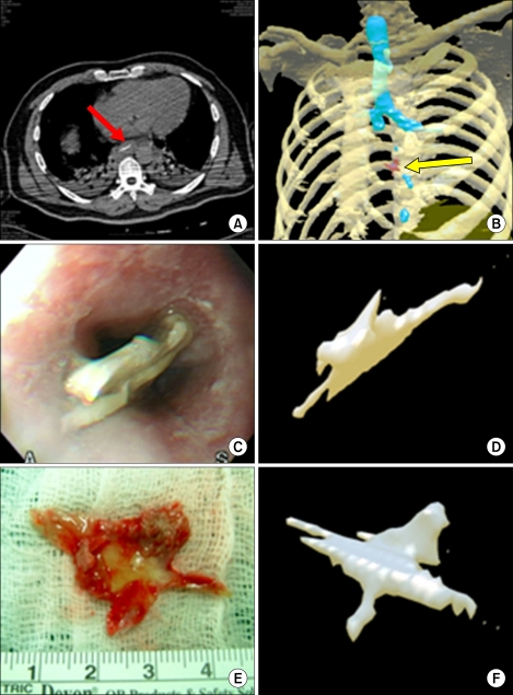
This study was conducted to investigate the clinical application of three-dimensional (3D) reconstructed computed tomography (CT) images in detecting and gaining information on esophageal foreign bodies (FBs). Two patients with esophageal FBs were enrolled for analysis. In both cases, 3D reconstructed images were compared with the FB that was removed according to the object shape, size, location, and orientation in the esophagus. The results indicate the usefulness of conversion of CT data to 3D images to help in diagnosis and treatment. Use of 3D images prior to treatment allows for rapid prototyping and surgery simulation.
Keywords: Computed tomography; Esophageal foreign body; Esophageal surgery.
Figures
References
-
- Young CA, Menias CO, Bhalla S, Prasad SR. CT features of esophageal emergencies. Radiographics. 2008;28:1541–1553. - PubMed
-
- Hirasaki S, Inoue A, Kubo M, Oshiro H. Esophageal large fish bone (Sea Bream Jawbone) impaction successfully managed with endoscopy and safely excreted through the intestinal tract. Intern Med. 2010;49:995–999. - PubMed
-
- Cheng HT, Wu CI, Tseng CS, et al. The occlusion-adjusted prefabricated 3D mirror image templates by computer simulation: the image-guided navigation system application in difficult cases of head and neck reconstruction. Ann Plast Surg. 2009;63:517–521. - PubMed
-
- Sannomiya EK, Silva JV, Brito AA, Saez DM, Angelieri F, Dalben Gda S. Surgical planning for resection of an ameloblastoma and reconstruction of the mandible using a selective laser sintering 3D biomodel. Oral Surg Oral Med Oral Pathol Oral Radiol Endod. 2008;106:e36–e40. - PubMed
Publication types
LinkOut - more resources
Full Text Sources