. 2012 Jan 20;14(1):R16.
doi: 10.1186/bcr3100.

STAT1-deficient mice spontaneously develop estrogen receptor α-positive luminal mammary carcinomas

Affiliations

STAT1-deficient mice spontaneously develop estrogen receptor α-positive luminal mammary carcinomas

Szeman Ruby Chan et al. Breast Cancer Res. .

Abstract

Introduction: Although breast cancers expressing estrogen receptor-α (ERα) and progesterone receptors (PR) are the most common form of mammary malignancy in humans, it has been difficult to develop a suitable mouse model showing similar steroid hormone responsiveness. STAT transcription factors play critical roles in mammary gland tumorigenesis, but the precise role of STAT1 remains unclear. Herein, we show that a subset of human breast cancers display reduced STAT1 expression and that mice lacking STAT1 surprisingly develop ERα+/PR+ mammary tumors.

Methods: We used a combination of approaches, including histological examination, gene targeted mice, gene expression analysis, tumor transplantaion, and immunophenotyping, to pursue this study.

Results: Forty-five percent (37/83) of human ERα+ and 22% (17/78) of ERα- breast cancers display undetectable or low levels of STAT1 expression in neoplastic cells. In contrast, STAT1 expression is elevated in epithelial cells of normal breast tissues adjacent to the malignant lesions, suggesting that STAT1 is selectively downregulated in the tumor cells during tumor progression. Interestingly, the expression levels of STAT1 in the tumor-infiltrating stromal cells remain elevated, indicating that single-cell resolution analysis of STAT1 level in primary breast cancer biopsies is necessary for accurate assessment. Female mice lacking functional STAT1 spontaneously develop mammary adenocarcinomas that comprise > 90% ERα+/PR+ tumor cells, and depend on estrogen for tumor engraftment and progression. Phenotypic marker analyses demonstrate that STAT1-/- mammary tumors arise from luminal epithelial cells, but not myoepithelial cells. In addition, the molecular signature of the STAT1-/- mammary tumors overlaps closely to that of human luminal breast cancers. Finally, introduction of wildtype STAT1, but not a STAT1 mutant lacking the critical Tyr701 residue, into STAT1-/- mammary tumor cells results in apoptosis, demonstrating that the tumor suppressor function of STAT1 is cell-autonomous and requires its transcriptional activity.

Conclusions: Our findings demonstrate that STAT1 suppresses mammary tumor formation and its expression is frequently lost during breast cancer progression. Spontaneous mammary tumors that develop in STAT1-/- mice closely recapitulate the progression, ovarian hormone responsiveness, and molecular characteristics of human luminal breast cancer, the most common subtype of human breast neoplasms, and thus represent a valuable platform for testing novel treatments and detection modalities.


Figures
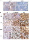
Figure 1
Figure 1
Immunohistochemical analysis of STAT1 expression levels in human breast cancers. (A) Immunohistochemical analysis on wild-type (WT) and STAT1-/- spleens to confirm the specificity of the antibody against STAT1. Sections of WT or STAT1-/- spleens were stained with a polyclonal rabbit antibody against an epitope in STAT1 which is homologous to both humans and mice. STAT1 reactivity was observed predominantly in the lymphoid cells of the white pulp in WT spleen. However, no appreciable signal was detected in the STAT1-/- spleen. Original magnification, 100×. Scale bars = 200 μm. (B) The staining intensity of the STAT1+ signal was evaluated by using a three-tiered scale (low, intermediate, and high). Low, intermediate, and high STAT1 correspond to the staining intensity scores of 1, 2, and 3, respectively, in Table 2. Six cases of estrogen receptor-alpha-negative (ERα-) and six cases of ERα+ primary human breast tumors are shown here as representative images. Original magnification, 200×. Scale bars = 100 μm.
Figure 2
Figure 2
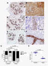
Selective downregulation of STAT1 expression in the neoplastic cells of human breast tumors. (A) Representative image of STAT1 staining on normal breast tissues of healthy individuals (n = 5) shows STAT1 expression in luminal epithelial cells and stromal cells and occasionally in myoepithelial cells. Original magnification, 100×. Scale bar = 200 μm. Inset, 400×. (B) Representative image of a breast tumor case with high levels of STAT1 expression in both the epithelial and stromal compartments. Original magnification, 200×. Scale bar = 100 μm. Inset, 400×. (C-F) Representative images of paired adjacent normal breast tissues (left) with breast tumors (right; n = 11). Morphologically normal breast tissues from patients with breast cancer display a STAT1 expression pattern that is similar to that in normal breasts from healthy individuals (C, E). Panel (C) is the paired normal tissue of the tumor in panel (D) (case 87), whereas panel (E) is the paired normal tissue of the tumor in panel (F) (case 58). The epithelial tumor cells of the representative estrogen receptor-alpha-positive (ERα+) (D) and ERα- (F) breast cancers were devoid of STAT1 expression, whereas STAT1 level remained high in the tumor stroma. Original magnification, 200×. Scale bars = 100 μm. (G) Intensity scores of the neoplastic and stromal cells of all cases (n = 161) are plotted. Reduced STAT1 staining intensity was preferentially associated with ERα+ human breast cancers relative to ERα- breast cancers in both the neoplastic and stromal compartments (P = 0.001, unpaired t test). Stromal cells had an overall higher STAT1 expression level than the neoplastic cells of the same tumors (P < 0.0001, Wilcoxon signed rank test). (H) Breast tumors exhibited significant reduction in STAT1 score in comparison with matched normal breast tissues (n = 11) (P = 0.003, Wilcoxon signed rank test). STAT1 score is the sum of the score representing the percentages of STAT1+ cells (percentage score) and that representing the STAT1 staining intensity (intensity score).
Figure 3
Figure 3
Spontaneous development of mammary gland adenocarcinomas in STAT1 deficient (STAT1-/-), STAT1 null (S1N), and STAT1-/- × RAG2-/- female mice. (A) STAT1-/- (n = 23, inverted red triangles), S1N (n = 26, inverted purple triangles), and STAT1-/- × RAG2-/- (n = 31, blue squares) mice succumbed to mammary tumors, whereas none of the age-matched wild-type (WT) 129S6/SvEv (n = 41, black triangles) or RAG2-/- (n = 33, yellow circles) mice developed the disease. P values were obtained with log-rank test by comparing STAT1-/- × RAG2-/- with WT mice (***P = 0.0003) and STAT1-/- or S1N with WT mice (****P < 0.0001). Curves between STAT1-/- and S1N mice are not different to a statistically significant degree (P = 0.24). (B) Multiparous STAT1-/- mice (n = 11, red) developed mammary tumors at a higher frequency and with shorter latency than nulliparous STAT1-/- mice (n = 12, black) (P < 0.0001).
Figure 4
Figure 4
Histopathological analyses of the mammary gland carcinomas developed in STAT1-/- mice. Histopathological analyses of the early lesions (A-H) and the invasive adenocarcinomas (I-N) in mammary glands of STAT1-/- mice. (A, B) Whole-mount images of a thoracic mammary fat pad show aberrant dilation of mammary ducts and clusters of cystic alveoli (arrow and arrowhead). Panel (B) is a magnified image of panel (A). Scale bars = 1 mm. (C-H) Histology of the cluster of cysts shown in panel (B), highlighted by an arrow in panel (B), demonstrated mammary intraepithelial neoplasia (MIN). (C, D, I, J) Sections were stained with hematoxylin and eosin (H&E). Solid nests of neoplastic cells with areas of necrosis and invasion are evident in the carcinomas (I, J). (E-H, K-N) Atypical cells in MIN lesion and neoplastic cells expressed estrogen receptor-alpha (ERα) (E, F, K, L) and progesterone receptor (PR) (G, H, M, N). (C, E, G, I, K, M) Scale bars = 100 μm. (D, F, H, J, L, N) Higher magnification of the corresponding left panels. Scale bars = 40 μm.
Figure 5
Figure 5
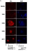
STAT1-/- mammary tumor cell lines SSM2 and SSM3 express estrogen receptor-alpha (ERα), PR-A, and PR-B. (A) SSM2 and SSM3 expressed nuclear ERα, similar to the human ERα+ breast cancer cell line MCF7. In contrast, NMuMG and SSM1 did not exhibit nuclear ERα staining. Scale bars = 40 μm. (B) SSM2 and SSM3 are positive for ERα expression, but SSM1 expressed a very low level of ERα, by Western blot analysis. NMuMG exhibited no detectable levels of ERα. (C) SSM2 and SSM3 expressed PR-A and PR-B, which are derived from alternative PR promoters and are target genes for ERα signaling, suggesting that SSM2 and SSM3 were estrogen-responsive. In contrast, NMuMG and SSM1 did not display functional ERα signaling. DAPI, 4'-6-diamidino-2-phenylindole; SSM, spontaneous STAT1-/- mammary (epithelial tumor cell line).
Figure 6
Figure 6
STAT1-/- mammary tumors respond to and depend on ovarian hormones for engraftment and tumor progression. (A) SSM1, SSM2, SSM3, or MCF7 was plated in phenol red-free media containing charcoal-treated fetal calf serum in the presence (17β-estradiol, or E2; black triangles) or absence (mock; red squares) of 10 nM E2. P values were obtained with t test comparing mock-treated and E2-treated samples. (B) SSM1, SSM2, or SSM3 (105 cells per mouse) was transplanted into the inguinal mammary glands of sham-treated (black triangles), ovariectomized (red squares), or ovariectomized wild-type or STAT1-/- mice that were supplemented with E2 pellets (blue circles). P values were obtained with t test comparing tumor growth in ovariectomized mice with that in either sham-treated or ovariectomized mice supplemented with E2 pellets [44]. (C) Growth of tumor transplants from primary STAT1-/- mammary tumors is ovarian hormone-dependent. Fragments (1 × 1 mm2) from two primary STAT1-/- mammary tumors were transplanted into the inguinal fat pads of ovariectomized mice (red) or sham-operated mice (black). No palpable masses were detected in ovariectomized mice. In contrast, tumors grew progressively in recipients with intact ovaries. Results represent seven to eight mice in each group (P < 0.0001). (D) SSM3 tumor cells were transplanted orthotopically into the mammary fat pads of nude mice. Mice bearing established tumors around 5 mm in diameter were sham-operated (black triangles; n = 7) or ovariectomized (red squares; n = 9). SSM3 tumors failed to progress in the absence of ovarian hormones, demonstrating sensitivity to ovarian hormone deprivation therapy (P < 0.0001). (E) Fragments of primary STAT1-/- mammary tumors were transplanted into the mammary fat pads of STAT1-/- mice. When the established tumors reached 5 mm in diameter, the animals were either sham-operated (black triangles; n = 3) or ovariectomized (red squares; n = 7). Transplanted primary tumors regressed after ovarian hormone deprivation (P < 0.0001). Error bars indicate standard error of the mean. OD540, optical density at 540 nm; OVX, ovariectomy; SSM, spontaneous STAT1-/- mammary (epithelial tumor cell line).
Figure 7
Figure 7
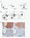
STAT1-/- mammary tumor cells display a luminal epithelial cell phenotype. (A) Gating procedure used for the analysis of cell surface markers. Disaggregated mammary glands or mammary tumors were collected by an LSRII flow cytometer and analyzed by using FlowJo. Cells were first selected on the basis of size by using forward and side scatter (left panel). Single cells were then selected by forward scatter (FSC)-A and FSC-W (middle panel). Live cells (DAPI-) and lineage- cells (Ter119-, CD31-, and CD45-) were gated for the analysis depicted in (B). (B) Expression of CD49f and CD24 on STAT1-/- mammary epithelial cells (MECs), mammary intraepithelial neoplasia (MIN), and carcinoma. Representative images from five STAT1-/- mice are shown. Myoepithelial (myo) and luminal epithelial (lum) are highlighted. (C) Immunohistochemical analysis of primary STAT1-/- mammary tumors for cytokeratin (CK) 5, 14, 8/18, and 19. Mammary tumor cells were stained positive for CK19 and CK8/18 (luminal epithelial markers) but negative for CK5 and CK14 (myoepithelial markers). Scale bar = 100 μm. DAPI, 4'-6-diamidino-2-phenylindole.
Figure 8
Figure 8
Gene expression profiles of primary STAT1-/- estrogen receptor-alpha-positive (ERα+) mammary tumors show significant overlap with that of human ERα+ luminal breast cancers. (A) Five primary STAT1-/- ERα+ mammary tumor datasets (S1_1, S1_2, S1_3, S1_4, and S1_6; orange) were analyzed with 232 annotated human breast cancer datasets and 13 other mouse mammary tumor models (purple) by hierarchical clustering using 96 genes known to be among those that are diagnostic for different human breast cancer subtypes [36]. Genes are represented in rows, and datasets are represented in columns. Red represents genes that are overexpressed compared with the median, whereas green represents genes that are underexpressed compared with the median. The STAT1-/- ERα+ mammary tumors (orange) cluster closely to the human luminal breast cancers (blue) at the far left of the heatmap, underscoring the significant relatedness between the two cohorts. All human samples are colored by intrinsic subtype as determined in [36]: blue = luminal, red = basal-like, pink = HER2+/ER-, yellow = claudin-low, and green = normal breast-like. Expression values of the datasets can be found in Additional file 3. (B) Display of seven genes that are important identifiers for the luminal subtype: KRT8, KRT18, XBP1, GATA3, MYB, AREG, and FOXA1 (from top to bottom). See Additional file 3 for the expression values of these genes. HER2, human epidermal growth factor receptor 2.
Figure 9
Figure 9
Tumor suppressor function of STAT1 is cell-autonomous. (A) STAT1 was ectopically expressed in SSM1, SSM2, and SSM3 by retroviral transduction (STAT1) to levels comparable to the endogenous level in nontransduced (-) NMuMG. STAT1 was also overexpressed in NMuMG (STAT1). Retrovirus expressing GFP alone was used as a negative control (GFP). (B) STAT1 reconstitution in SSM2 and SSM3 resulted in 4.5- and 3.5-fold increases in early apoptosis (that is, annexin V-positive, 7-AAD-negative cells) 3 days post-transduction, respectively. **P < 0.005. (C) Tyrosine 701 is required for STAT1-mediated apoptosis in SSM2 and SSM3. Retrovirus expressing GFP, WT STAT1 (STAT1), and STAT1 mutants (Y701F or S727A) were transduced into SSM2 and SSM3. Mutation in Y701 (Y701F) abolished the ability of STAT1 to induce cell death, whereas that in S727 (S727A) was still capable of inducing cell death. SSM2, *P = 0.02. SSM3, *P = 0.04. P values were obtained with unpaired t test. ns, not significant; SSM, spontaneous STAT1-/- mammary (epithelial tumor cell line).

Similar articles

Cited by

References

    1. Anderson WF, Chatterjee N, Ershler WB, Brawley OW. Estrogen receptor breast cancer phenotypes in the surveillance, epidemiology, and end results database. Breast Cancer Res Treat. 2002;76:27–36. doi: 10.1023/A:1020299707510. - DOI - PubMed
    1. Bundred NJ. Prognostic and predictive factors in breast cancer. Cancer Treat Rev. 2001;27:137–142. doi: 10.1053/ctrv.2000.0207. - DOI - PubMed
    1. Perou CM, Sorlie T, Eisen MB, van de Rijn M, Jeffrey SS, Rees CA, Pollack JR, Ross DT, Johnsen H, Akslen LA, Fluge O, Pergamenschikov A, Williams C, Zhu SX, Lonning PE, Borresen-Dale AL, Brown PO, Botstein D. Molecular portraits of human breast tumours. Nature. 2000;406:747–752. doi: 10.1038/35021093. - DOI - PubMed
    1. Chia YH, Ellis MJ, Ma CX. Neoadjuvant endocrine therapy in primary breast cancer: indications and use as a research tool. Br J Cancer. 2010;103:759–764. doi: 10.1038/sj.bjc.6605845. - DOI - PMC - PubMed
    1. Simian M, Manzur T, Rodriguez V, de Kier Joffe EB, Klein S. A spontaneous estrogen dependent, tamoxifen sensitive mouse mammary tumor: a new model system to study hormone-responsiveness in immune competent mice. Breast Cancer Res Treat. 2009;113:1–8. doi: 10.1007/s10549-007-9888-x. - DOI - PubMed

Publication types

MeSH terms