. 2012 Feb;93(1):1-10.
doi: 10.1111/j.1365-2613.2011.00807.x.

Fell-Muir Lecture: chondroitin sulphate glycosaminoglycans: fun for some and confusion for others

Affiliations

Fell-Muir Lecture: chondroitin sulphate glycosaminoglycans: fun for some and confusion for others

Bruce Caterson. Int J Exp Pathol. 2012 Feb.

Abstract

This review emphasizes the importance of glycobiology in nature and aims to highlight, simplify and summarize the multiple functions and structural complexities of the different oligosaccharide combinatorial domains that are found in chondroitin sulphate/dermatan sulphate (CS/DS) glycosaminoglycan (GAG) chains. For example, there are 1008 different pentasaccharide sequences possible within CS, DS or CS/DS hybrid GAG chains. These combinatorial possibilities provide numerous potential ligand-binding domains that are important for cell and extracellular matrix interactions as well as specific associations with cytokines, chemokines, morphogens and growth factors that regulate cellular differentiation and proliferation during tissue development, for example, morphogen gradient establishment. The review provides some details of the large and diverse number of different enzymes that are involved in CS/DS biosynthesis and attempts to explain how differences in their expression patterns in different cell types can lead to subtle but important differences in the GAG metabolism that influence cellular proliferation and differentiation in development as well as regeneration and repair in disease. Our laboratory was the first to generate and characterize monoclonal antibodies (mAb) that very specifically recognize different ‘native’ sulphation motif/epitopes in CS/DS GAG chains. These monoclonal antibodies have been used to identify very specific spatio-temporal expression patterns of CS/DS sulphation motifs that occur during tissue and organ development (in particular their association with stem/progenitor cell niches) and also their recapitulated expression in adult tissues with the onset of degenerative joint diseases. In summary, diversity in CS/DS sulphation motif expression is a very important necessity for animal life as we know it.


Figures

Figure 1
Figure 1
Structural Diversity of Glycosaminoglycan repeating disaccharide units. The repeating disaccharide units of Hyaluronan (HA), Keratan Sulphate (KS), Chondroitin Sulphate (CS), Dermatan Sulphate (DS), Heparin (Hep) and Heparan Sulphate (HS) consist of a hexosamine [either Glucosamine (GlcN) or Galactosamine (GalN) - Squares] and a uronic acid [either Glucuronic (GlcUA) or Iduronic acid (IdUA) - Triangles] or Galactose (Gal) –Circles. These sugar and disaccharide units are joined to one another by different anomeric glycosidic linkages (α1-4, β1-3 or β1-4) between the sugar hydroxyl groups. The hexosamine, uronic acid and galactose sugars in each disaccharide unit can also be substituted with sulphate groups on the sugar hydroxyls in the 2- (2S), 3- (3S), 4- (4S) and 6- (6S) positions. The hexosamine is usually N-acetlyated (NAc), but in Hep/HS, it can also be N-sulphated (NS) or occur as the free amine (N). HA is the only non-sulphated GAG and it is also not found covalently attached to a proteoglycan core protein.
Figure 2
Figure 2
Structure and location of native epitopes [for mAbs 3-B-3(−), 4-C-3, 6-C-3 and 7-D-4] and chondroitinase-generated ‘stub’ neoepitopes [for mAbs 1-B-5, 2-B-6 and 3-B-3(+)] on chondroitin sulphate (CS) and dermatan sulphate (DS) glycosaminoglycan chains. Boxed in Red is the location of mAb 3-B-3(−) native epitope that recognizes a non-reducing terminal end saturated CS disaccharide consisting of glucuronic acid that is adjacent to N-acetylgalactosamine-6-sulphate. The native epitope for mAb 6-C-3 (Red linear arrows) is located in CS/DS oligosaccharide sequences that are located on the outer peripheral regions of the CS/DS GAG chains whilst mAbs 4-C-3 and 7-D-4 are located in oligosaccharide sequences located in the inner regions of the CS/DS GAG chains nearer to the linkage region (LR) that is the covalent attachment site that links CS/DS GAGs to the proteoglycan core protein. The chondroitinase-generated neoepitope ‘CS stubs’ are circled in Red and occur as an unsaturated uronic acid residue that is adjacent to N-acetylgalactosamine that is either non-sulphated (C-O-S; recognized by mAb 1-B-5), 4-sulphated (C-4-S; recognized by mAb 2-B-6) or 6-sulphated [C-6-S; recognized by mAb 3-B-3(+)]. CS/DS GAG chains are depicted as a sequence of disaccharides with two different coloured boxes that have either been partially (limited) or completely digested (deglycosylated) with chondroitinase. Chodroitinases are enzymes that remove CS/DS disaccharides with non-reducing unsaturated uronic acid residues from the CS/DS GAG chains eventually leaving a characteristic terminal disaccharide ‘stub’, that is, either unsulphated, 4- or 6-sulphated, and covalently attached to the proteoglycan core protein.
Figure 3
Figure 3
Specific spatial and temporal expression of native CS/DS sulphation motif epitopes recognized by mAbs 3B3(−) and 6C3 in developing embryonic chick tissues at stages E12 and E14. Joint tissues from stage E12 [A–F] and E14 [G–L] embryonic chicks stained with Alcian Blue [A,B,G,H] and immunostaining with mAbs 3B3(−) [C,D,I,J] and 6C3 [E,F,K,L]. Circles indicate regions shown at higher magnifications in adjacent frames. The asterisks (*) highlight unstained fibrocartilagenous articular cartilage; that is, at E12 mAb 6C3 is broadly labelling the epiphyses and not the overlaying fibrous articular cartilage, whereas 3B3(−) immunostaining is different and identifies a specific subset of cells and matrix in the developing articular cartilage.
Figure 4
Figure 4
Specific spatial expression of native CS/DS sulphation motif epitopes recognized by mAbs 3B3(−), 4C3 and 7D4 in a 12-week-old human foetal knee joint. Sections have been stained with Toluidine Blue [A] and immunostained with 3B3(−) [B], 4C3 [C] and 7D4 [D]. Note that Toluidine Blue staining identifies the presence of the major populations of CS GAG chains in the developing knee joint tissues. This Toluidine Blue staining is widespread and identifies the large number of GAGs present (mainly CS) throughout the early cartilage elements of the femur (f) and tibia (t) with weaker staining of the fibrocartilagenous meniscus (m) and other fibrous connective tissues of the joint. In contrast, immunostaining with the mAbs 3B3(−), 4C3 and 7D4 shows positive staining in very specific zones of the developing joint cartilage and meniscus that clearly delineate where the future articular cartilage and inner portions of the meniscus will eventually be. Close inspection of the three different mAb staining patterns shows that there are subtle differences in the mAb staining in the matrix or on the cells themselves indicating the specific spatial expression of these CS/DS sulphation epitopes.
Figure 5
Figure 5
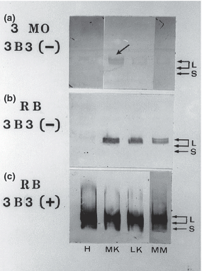
Agarose-acrylamide gel separation and Western blot analyses using mAb 3B3(−) and (+) of proteoglycan subpopulations extracted from the Hip (H) and Knee Joint cartilages (i.e. MK – medial; LK – lateral and MM – medial meniscus cartilages) of 3 month old (A) and 9 month old (B & C) retired breeder (RB) Hartley strain Guinea Pigs. Large aggregating proteoglycans (L – i.e. aggrecan subpopulations) and small leucine-rich proteoglycans (S) are identified by mAb 3B3 immunostaining either on the untreated nitrocellulose membranes [A & B where native 3B3(−) CS GAG chain epitopes are present – see Figure 2] or after the nitrocellulose membranes have been subjected to a chondroitinase ABC digestion [C] prior to their immunostaining with 3B3(+) where ‘CS-stubs’ containing a terminal unsaturated uronic acid adjacent to galactosamine-6-sulphate are present on both L and S proteoglycan subpopulations [C]. Note: only the slowest migrating (newly synthesized) aggrecan subpopulation is identified by immunostaining with 3B3(−) in [A] and [B]. The arrow points to the 3B3(−) positive staining that can only be found in the medial knee cartilage of 3-month-old animals.
Figure 6
Figure 6
The Glycosaminoglycan Poem. Inspired from the poem ‘My Country’ (2nd verse) composed by Dorothea MacKeller OBE (1885–1968).

Similar articles

Cited by

References

    1. Bautch J, Clayton M, Chu Q, Johnson K. Synovial fluid chondroitin sulphate epitopes 3B3 and 7D4, and glycosaminoglycan in human knee osteoarthritis after exercise. Ann. Rheum. Dis. 2000;59:887–891. - PMC - PubMed
    1. Benjamin AL, Rapraeger AC. Spacial and temporal expression of heparan sulphate in mouse development regulates FGF and FGF receptor assembly. J. Cell Biol. 2003;163:637–648. - PMC - PubMed
    1. Carlson CS, Loeser RF, Johnstone B, Tulli HM, Dobson DB, Caterson B. Osteoarthritis om cynomolgus,acaques. II. Detection of modulated proteoglycan epitopes in cartilage and synovial fluid. J. Orthop. Res. 1995;13:399–409. - PubMed
    1. Carney SL, Billingham MEJ, Caterson B, et al. Changes in proteoglycan turnover in experimental canine osteoarthritic cartilage. Matrix. 1992;12:137–147. - PubMed
    1. Caterson B, Christner JE, Baker JR. Characterization of a monoclonal antibody that specifically recognizes Corneal and Skeletal Keratan Sulfate. J. Biol. Chem. 1983;258:8848–8854. - PubMed

Publication types

MeSH terms

LinkOut - more resources