. 2012 Jan 20:9:6.
doi: 10.1186/1477-7517-9-6.

Injecting drug use via femoral vein puncture: preliminary findings of a point-of-care ultrasound service for opioid-dependent groin injectors in treatment

Affiliations

Injecting drug use via femoral vein puncture: preliminary findings of a point-of-care ultrasound service for opioid-dependent groin injectors in treatment

Richard Senbanjo et al. Harm Reduct J. .

Abstract

Background: Within the UK, injecting in the femoral vein (FV), often called 'groin injecting', is a serious cause of risk and harm. This study aimed to use ultrasound scanning as a means to engage groin injectors (GIs), examine their femoral injecting sites and assess their venous health, with the intention of developing improved responses.

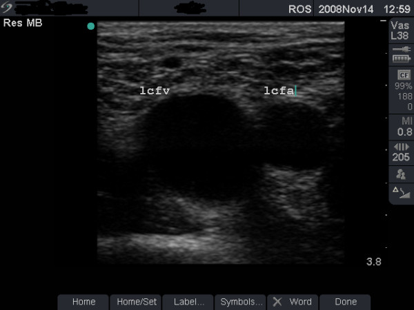
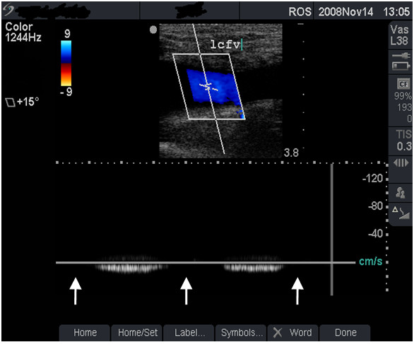
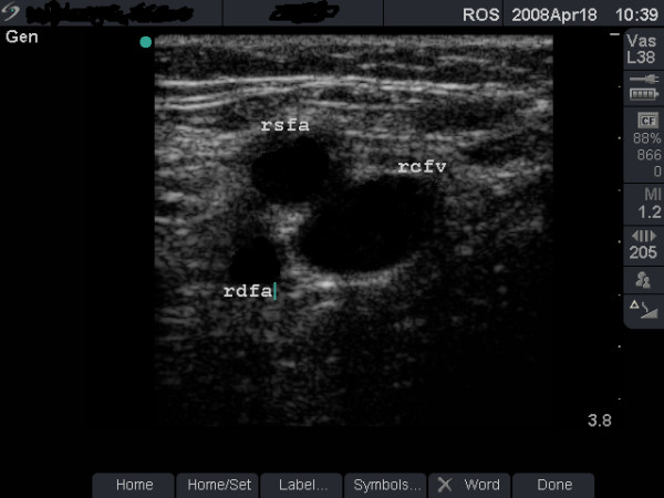
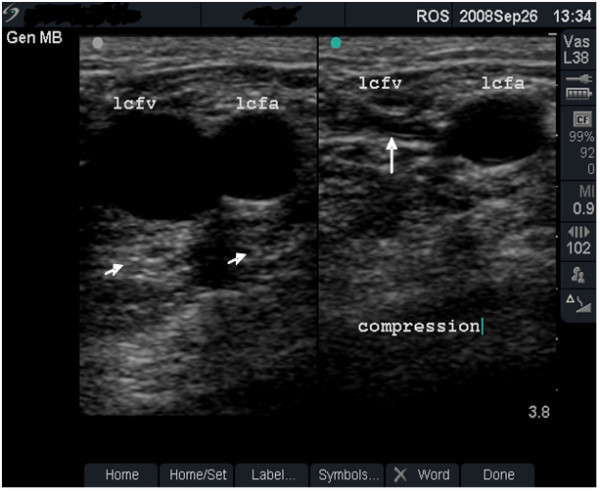
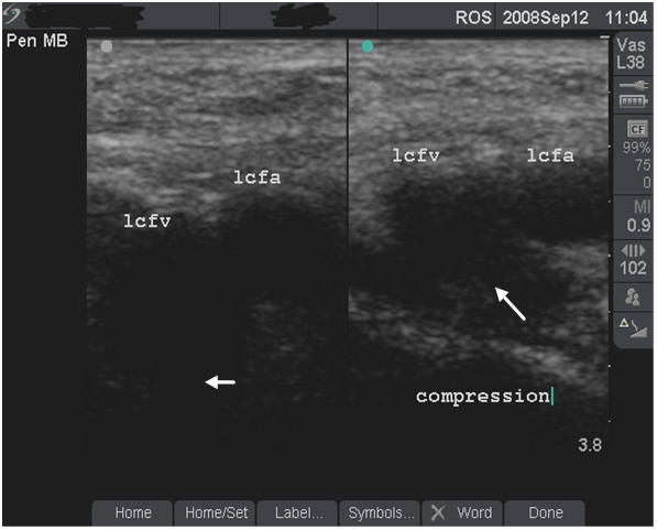
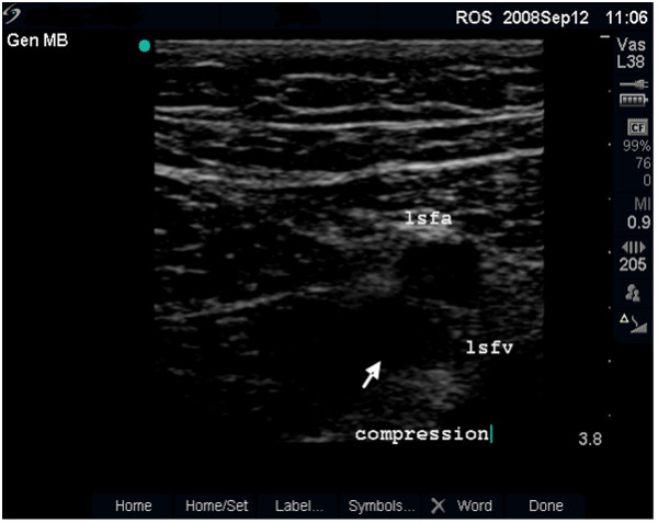
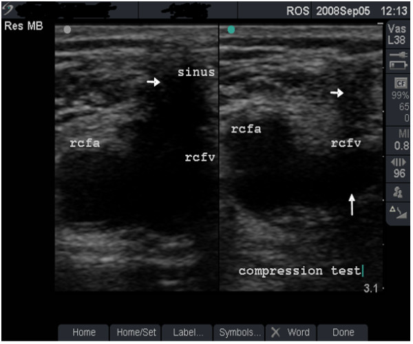
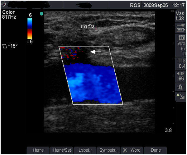
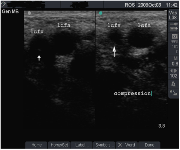
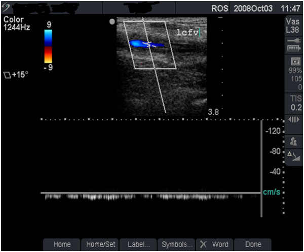
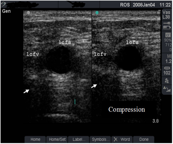
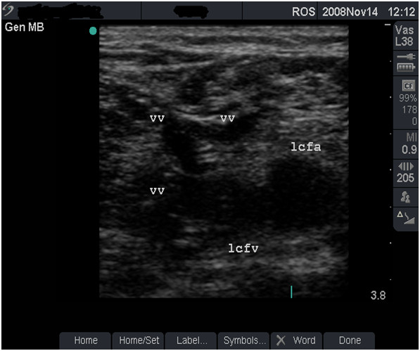
Methods: Between September 2006 and March 2009, GIs attending a network of community drug treatment centres in South East England were invited to attend an ultrasound 'health-check' clinic. This paper provides a narrative account of the scanning procedure and operation of the service, with descriptive statistical analysis of GIs who attended. The analysis uses a structured, specially-developed clinical data set that incorporates a categorisation for the severity of FV damage. Case studies using ultrasound images and a link to a video are provided to illustrate the range of presentations encountered and the categorisations used for severity.

Results: A total of 160 groin scans (76 bilateral and 8 unilateral) were performed in 84 GIs. The majority were men (69.0%) and the mean age of the sample was 36.8 years. The mean duration of drug use and injecting drug use was 19.7 years and 13.8 years, respectively. FV damage at the injecting site in the right groin was graded as minimal in 20 patients (25%), moderate in 27 (33.8%), severe in 16 (20.0%) and very-severe in 17 (21.3%). Corresponding figures for left FV were 24 (30.0%), 22 (27.5%), 18 (22.5%) and 16 (20.0%). Wide variation was observed in the time to the development of these grades of FV damage.

Conclusions: Modern, portable ultrasound scanners make it possible to examine the venous health of GIs in community treatment settings. Ultrasound scanning identified extensive FV damage, much hitherto-unrecognised in this population. These findings should further alert clinicians, policy-makers and patients to the urgent need for effective harm reduction responses to GI behaviour. Images of damaged FV in this paper might prove to be a useful resource for discussions about GI risks.


Figures

Figure 1
Figure 1
Vein with minimal damage (grade 1). B-mode image of the left femoral vein (lcfv) and artery (lcfa) in a 30-year old groin injector (case study B). The damage to this vein is minimal (grade 1).
Figure 2
Figure 2
Normal venous flow (grade 1). Duplex scan showing normal blood flow - colour flow (top half) and spectral tracing (lower half) in the left femoral vein (lcfv) of the patient in figure 1 (case study B). Note the normal, phasic venous flow with respiration (arrows).
Figure 3
Figure 3
Femoral artery anterior to femoral vein. B-mode image taken 3 cm below the right inguinal ligament in a 24-year old groin injector. Note that the femoral artery has divided into the femoral artery of the thigh (rsfa) and the profundus (deep) artery of the thigh (rdfa).
Figure 4
Figure 4
Fully compressible, normal vein (grade 1). Dual-frame image of the left femoral vein (lcfv) and artery (lcfa) in a 44-year old groin injector who has never injected in the lcfv. Patency of the lcfv is demonstrated by the complete collapse of the vein with transducer pressure (long arrow). Note the post-acoustic shadows (short arrows).
Figure 5
Figure 5
Non-compressible vein (dual frame). Dual-frame image of the left femoral vein (lcfv) and artery (lcfa) in a 31-year old woman (case study A). The lcfv is non-compressible (long arrow) suggestive of acute deep vein thrombosis (DVT). Note the enhanced echo pattern in the soft tissue caused by oedema and the absence of post-acoustic shadow (small arrow).
Figure 6
Figure 6
Non-compressible vein (B mode). B-mode image of a non-compressible left femoral vein (lsfv, see arrow) behind the femoral artery (lsfa) in the thigh of the patient in figure 5 (case study A), indicative of extension of the blood clot to the femoral vein in the mid-thigh.
Figure 7
Figure 7
Vein with moderate damage (grade 2). Dual-frame image and compression test in a 36-year old man (case study C). A sinus (short arrows) is seen above the right femoral vein (rcfv) which is not fully compressible (long arrow). This vein damage was graded as moderate (grade 2).
Figure 8
Figure 8
Venous flow with minor abnormality (grade 2). Colour flow image from the right femoral vein (rcfv) of the patient in figure 7 (case study C). Note that venous blood flow (as indicated in colour flow image) beneath the sinus remains satisfactory (arrow).
Figure 9
Figure 9
Vein with severe damage (grade 3). Dual-frame image and compression test in a 43-year old woman (case study D). The left femoral vein (lcfv, short arrow) is smaller than the artery (lcfa) and the vein did not collapse completely with compression (long arrow). The damage to this vein was graded as severe (grade 3).
Figure 10
Figure 10
Vein with reduced, continuous flow (grade 3). Duplex scan showing significant reduction in blood flow (top half of image) and continuous venous flow (lower half of image) in the severely damaged left femoral vein (lcfv) of the patient in figure 9 (case study D). Compare this reduced, continuous flow with the normal venous flow pattern in figure 2.
Figure 11
Figure 11
No blood flow, very severe damage (grade 4). Dual-frame image from a 37-year old groin injector (see case study E). The left femoral vein (lcfv) is small and occluded by an echogenic material (organised blood clot). Note the absence of post-acoustic shadow behind the vein (arrows). The damage was graded as very-severe (grade 4).
Figure 12
Figure 12
Alternative venous drainage, very severe damage (grade 4). B-mode image showing a very-severely damaged left femoral vein (lcfv) and new vessels (vv), providing alternative routes for venous drainage.

Similar articles

Cited by

References

    1. Hay G, Gannon M, MacDougall J, Millar T, Eastwood C, McKeganey N, L G. In: Measuring different aspects of problem drug use: Methodological developments. Singleton N, Murray R, L T, editor. London: Home Office; 2006. Local and national estimates of the prevalence of opiate use and/or crack cocaine use (2004/05)
    1. De Angelis D, Hickman M, Yang SY. Estimating long-term trends in the incidence and prevalence of opiate use/injecting drug use and the number of former users: Back-calculation methods and opiate overdose deaths. Am J Epidemiol. 2004;160:994–1004. doi: 10.1093/aje/kwh306. - DOI - PubMed
    1. Maliphant J, Scott J. Use of the femoral vein ('groin injecting') by a sample of needle exchange clients in Bristol, UK. Harm Reduction Journal. 2005;2:6. doi: 10.1186/1477-7517-2-6. - DOI - PMC - PubMed
    1. Rhodes T, Stoneman A, Hope V, Hunt N, Martin A, Judd A. Groin injecting in the context of crack cocaine and homelessness: From 'risk boundary' to 'acceptable risk'? Int J Drug Policy. 2006;17:164–170. doi: 10.1016/j.drugpo.2006.02.011. - DOI
    1. Williams K, Abbey E. Knowledge of deep vein thrombosis among intravenous drug misusers. Psychiatric Bulletin. 2006;30:263–265. doi: 10.1192/pb.30.7.263. - DOI

LinkOut - more resources