. 2012 Jan 20:7:4.
doi: 10.1186/1749-8104-7-4.

Sonic hedgehog lineage in the mouse hypothalamus: from progenitor domains to hypothalamic regions

Affiliations

Sonic hedgehog lineage in the mouse hypothalamus: from progenitor domains to hypothalamic regions

Gonzalo Alvarez-Bolado et al. Neural Dev. .

Abstract

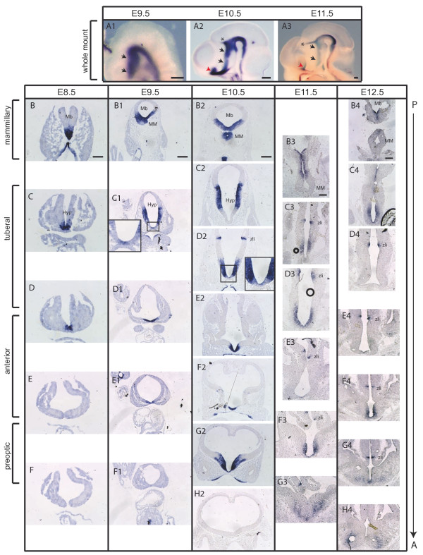
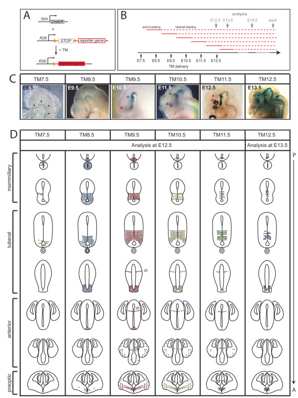
Background: The hypothalamus is a brain region with essential functions for homeostasis and energy metabolism, and alterations of its development can contribute to pathological conditions in the adult, like hypertension, diabetes or obesity. However, due to the anatomical complexity of the hypothalamus, its development is not well understood. Sonic hedgehog (Shh) is a key developmental regulator gene expressed in a dynamic pattern in hypothalamic progenitor cells. To obtain insight into hypothalamic organization, we used genetic inducible fate mapping (GIFM) to map the lineages derived from Shh-expressing progenitor domains onto the four rostrocaudally arranged hypothalamic regions: preoptic, anterior, tuberal and mammillary.

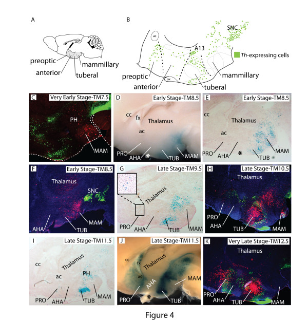
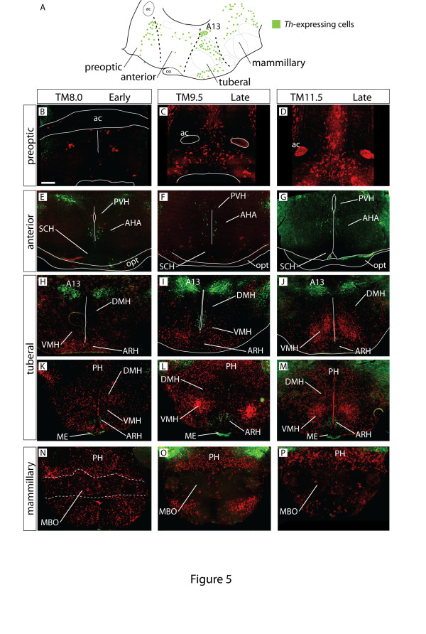
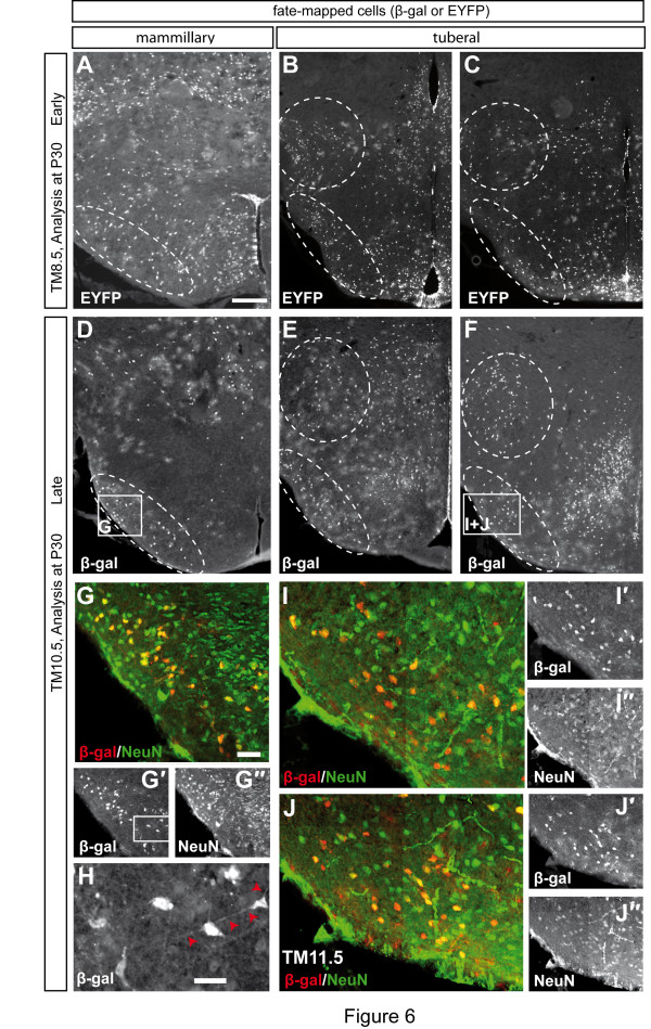
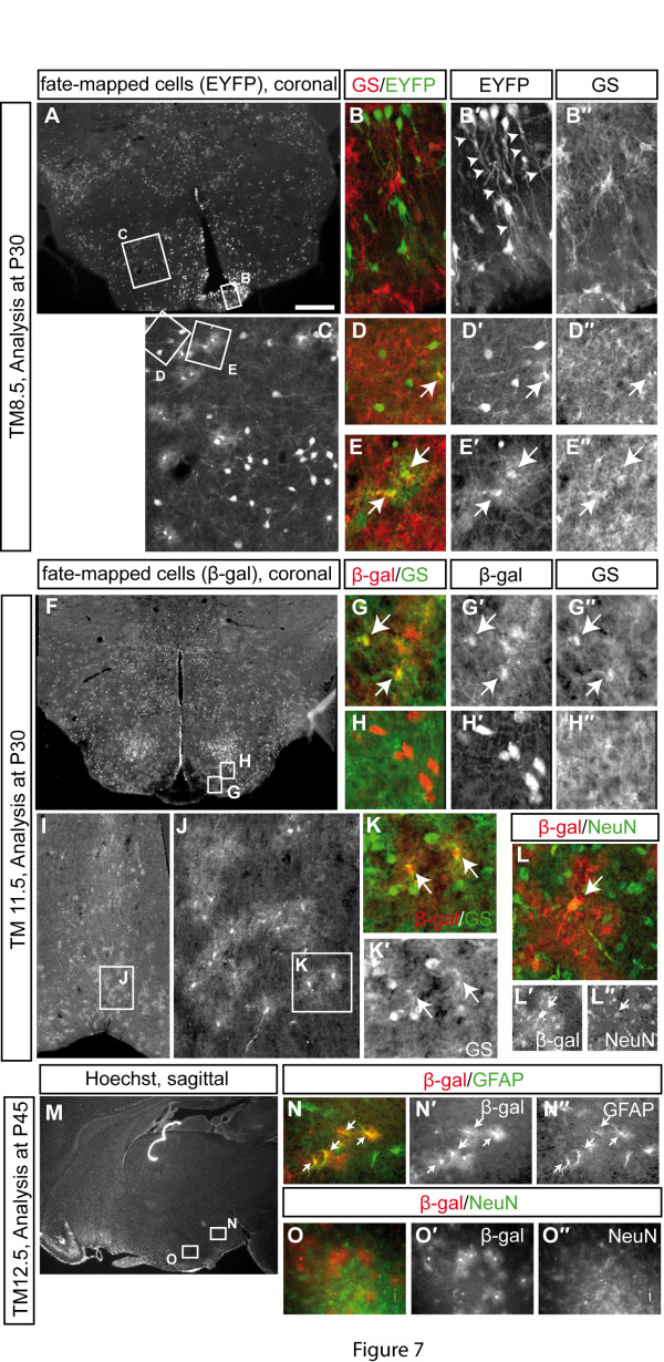
Results: Shh-expressing progenitors labeled at an early stage (before embryonic day (E)9.5) contribute neurons and astrocytes to a large caudal area including the mammillary and posterior tuberal regions as well as tanycytes (specialized median eminence glia). Progenitors labeled at later stages (after E9.5) give rise to neurons and astrocytes of the entire tuberal region and in particular the ventromedial nucleus, but not to cells in the mammillary region and median eminence. At this stage, an additional Shh-expressing domain appears in the preoptic area and contributes mostly astrocytes to the hypothalamus. Shh-expressing progenitors do not contribute to the anterior region at any stage. Finally, we show a gradual shift from neurogenesis to gliogenesis, so that progenitors expressing Shh after E12.5 generate almost exclusively hypothalamic astrocytes.

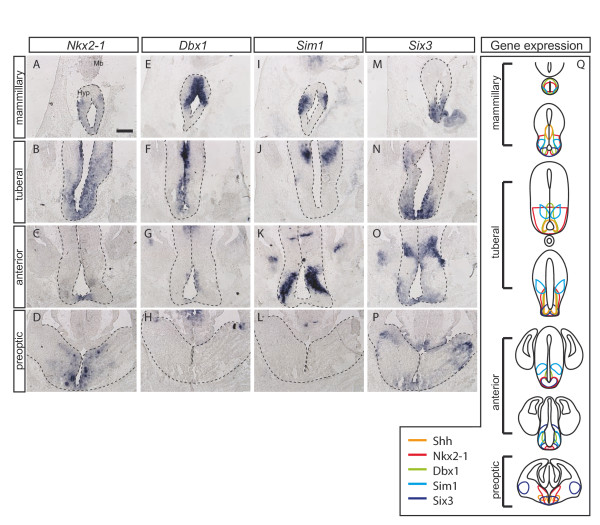
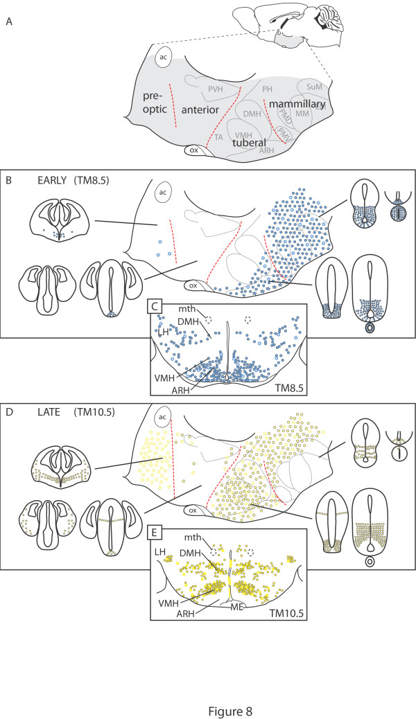
Conclusions: We define a fate map of the hypothalamus, based on the dynamic expression of Shh in the hypothalamic progenitor zones. We provide evidence that the large neurogenic Shh-expressing progenitor domains of the ventral diencephalon are continuous with those of the midbrain. We demonstrate that the four classical transverse zones of the hypothalamus have clearly defined progenitor domains and that there is little or no cell mixing between the tuberal and anterior or the preoptic and anterior hypothalamus. Finally, we show that, in the tuberal hypothalamus, neurons destined for every mediolateral level are produced during a period of days, in conflict with the current 'three-wave' model of hypothalamic neurogenesis. Our work sets the stage for a deeper developmental analysis of this complex and important brain region.


Figures

Figure 1
Figure 1
The dynamic expression pattern of Shh in the hypothalamic primordium. (A-H) RNA in situ hybridization in whole mounts (A1-A3) or on transverse sections of the hypothalamic primordium (hyp) at several anterior-posterior levels (B-H4). Shh is initially expressed in the ventral midline in the posterior hyp at E8.5 (black arrows in A1-A3; B-F). Shh expression expands laterally over the subsequent days of development and starts to be downregulated in the ventral midline (E9.5 to E12.5) (A1-H4). At E9.5 to E12.5 Shh is also expressed in the preoptic area and the zona limitans intrathalamica (zli). Red arrowheads in (A2, A3) indicate the preoptic area, and asterisks indicate the zli. Insets in (C1, D2): higher magnifications of the boxed area. Mb, midbrain; MM, mammillary primordium. Scale bars: 80 μm (B-F); 200 μm (A1-A3, B1-H4).
Figure 2
Figure 2
Changing populations of precursors are marked with Shh-GIFM at different stages of development. (A) Alleles used for GIFM. ShhCreER mice were crossed with R26 reporter mice. The reporter gene is either lacZ or EYFP. Administration of tamoxifen (TM) to pregnant females results in Cre-mediated recombination of the reporter allele and permanent expression of the reporter gene. (B) Since TM is only stable for 24 to 36 hours, the period of Cre-mediated recombination is restricted to approximately one day after TM administration (active labeling). Labeling is retained (retained labeling) after Cre activity ceases (due to degradation of TM or downregulation of Cre expression). The retained labeling allows for the fate-mapping of recombined and marked cells. (C) X-gal whole mount staining of ShhCreER/+; R26lz/+ embryos one day after the indicated time point of TM administration. (We refer to the time point of TM injection as TM followed by the embryonic day - for example, TM administration at E7.5 is TM7.5, and so on.) Black arrowheads indicate the notochord/prechordal plate, red arrowheads indicate the preoptic area, asterisks indicate the zona limitans intrathalamica (zli). (D) Summary of the distribution of Shh-expressing cells that were permanently marked at the indicated time points (TM7.5 to TM12.5) and analyzed at E12.5 or E13.5. Cells derived from Shh-expressing progenitors are initially found in the ventral midline of the posterior hypothalamic primordium (TM7.5 and TM8.5), but are later (TM9.5 to TM12.5) excluded from the midline. Shh-derived cells are also found in the preoptic area and the zli at later stages, but are almost completely excluded from the anterior hypothalamus.
Figure 3
Figure 3
Transcription factor domains in the developing hypothalamus. (A-P) RNA in situ hybridization on E12.5 transverse sections. The ventral diencephalon of selected sections along the anterior-posterior axis is shown. Hyp, hypothalamus; Mb, midbrain. (Q) Summary of gene expression domains. Note that only gene expression domains in the hypothalamus are indicated in the schematic, and thalamic gene expression is not indicated. Scale bar: 200 μm.
Figure 4
Figure 4
Two discrete domains of Shh-lineage along the rostrocaudal hypothalamic axis. (A) Adult mouse brain in sagittal view showing the hypothalamus (shaded gray) and its four transverse subdivisions. (B) Schematic distribution of tyrosine hydroxylase (Th) expressing cells (green dots) on a sagittal view of the hypothalamus (with information from the Allen Brain Atlas [33]). (C-K) Brains of ShhCreER/+; R26lz/+ mice. Tamoxifen (TM) was administered as indicated and sections or whole-mount brains were labeled to detect the Shh-lineage. (C, F, H, K) Sagittal sections of adult hypothalamus labeled for Shh lineage (β-gal, red) and Th (green), TM as indicated. Dashed line outlines the brain. (D, J) Right side of hemi-dissected adult brain labeled for Shh-lineage (X-gal reaction, blue), TM as indicated. (E, G, I) Sagittal sections through the E18.5 hypothalamus labeled for Shh lineage (X-gal reaction, blue), TM as indicated. (G) Only few fate-mapped cells are found in the preoptic area with TM9.5. The boxed area is shown in higher magnification. Asterisks indicate the optic chiasm.A13, dopaminergic cell group A13; ac, anterior commissure; AHA, anterior hypothalamic region; cc, corpus callosum; fx, fornix; MAM, mammillary region; Ox, optic chiasm; PH, posterior hypothalamus; PRO, preoptic region; SNC, substantia nigra pars compacta; TUB, tuberal region.
Figure 5
Figure 5
Preoptic glia and most tuberal and mammillary neurons are of Shh descent. (A) Schematic distribution of tyrosine hydroxylase (Th)-expressing cells (green dots) on a sagittal view of the hypothalamus. (B-P) Transverse sections of adult mouse brains TM8.0 (B, E, H, K, N), TM9.5 (C, F, I, L, O) and TM11.5 (D, G, J, M, P) labeled for Shh lineage (β-gal, red) and Th (green). (B-D) The preoptic region contains large numbers of late-stage fate-mapped cells with glia-like morphology. (E-G) The anterior hypothalamus contains very few fate-mapped cells at all stages. (H-M) Many fate-mapped cells are located in the tuberal area at all stages, especially in the ventromedial nucleus (VMH). (N-P) Early-stage fate-mapped cells (TM8.5) also contribute strongly to the mammillary body. A13, dopaminergic cell group A13; ac, anterior commissure; AHA, anterior hypothalamic region; ARH, arcuate nucleus; DMH, dorsomedial nucleus; opt, optic tract; MBO, mammillary body; ME, median eminence; Ox, optic chiasm; PH, posterior hypothalamus; PVH, paraventricular hypothalamic nucleus; SCH, suprachiasmatic nucleus; VMH, ventromedial nucleus. Scale bar: 800 μm.
Figure 6
Figure 6
Hypothalamic neurons derived from progenitors that express Shh after E10.5 settle in medial and lateral aspects of the tuberal hypothalamus. (A-F) Early and late stage fate-mapped cells contribute to the lateral tuberal and mammillary hypothalamus. Ventro- and dorsolateral areas of the hypothalamus are circled with a dashed line. (G-I) Immunostaining for fate-mapped cells (β-gal, red) and NeuN, a pan-neuronal marker (green). Pictures were taken using a Zeiss Apotome set-up. Late-stage fate-mapped cells overlap with the neuronal marker NeuN and have neuronal morphology (H). Red arrowheads indicate neuronal processes. Scale bar: 800 μm (A-F); 100 μm (G, I, J); 20 μm (H).
Figure 7
Figure 7
Shh-expressing progenitors give rise to astrocytes in the hypothalamus. Immunostaining for glutamin synthetase (GS) and fate-mapped cells (β-gal or EYFP) (B, D, E, G, H, K), GFAP and β-gal (N) or NeuN and β-gal (L, O). Sections were imaged using a Zeiss Apotome setup. (A, B) In the tuberal hypothalamus, fate-mapped cells (TM8.5, green) are localized to the median eminence and display the typical morphology of tanycytes (arrowheads). (C-H) Tuberal hypothalamus. Fate-mapped cells with astrocytic morphology overlap with GS (arrows), and marked cells with neuronal morphology do not express GS. (I-K) Almost all fate-mapped cells in the preoptic area have astrocytic morphology and overlap with GS. (L) Only few fate-mapped cells in the preoptic area overlap with the neuronal marker NeuN. (M-O) Progenitors expressing Shh after E12.5 give rise to astrocytes but not neurons. Arrows indicate double-labeled cells. Scale bars: 800 μm (A, F, I); 100 μm (C, J, N, O); 40 μm (B, D, E, G, H, K, L). P, postnatal day.
Figure 8
Figure 8
Distribution of Shh-lineage cells from the different progenitor domains in the mouse adult hypothalamus. (A) Adult mouse brain in sagittal view showing the hypothalamus (shaded gray). (B, D) Summary of distribution of cells fate-mapped at early and late stages. Magnified view of the shaded area showing the transverse hypothalamic subdivisions and some of the largest nuclei and Shh-lineage progenitor domains in the E12.5 neuroepithelium (small profiles; see also Figure 2). In the schematic of the adult hypothalamus, closed circles indicate neurons, open circles indicate astrocytes, and stars indicate tanycytes. (C, E) Transverse section through the adult tuberal hypothalamus. ac, anterior commissure; ARH, arcuate nucleus; DMH, dorsomedial nucleus; LH, lateral hypothalamus; LM, lateral mammillary nucleus; ME, median eminence; MM, medial portion of mammillary body; mth, mammillothalamic tract; ox, optic chiasm; PH, posterior hypothalamus; PMD, dorsal premammillary nucleus; PMV, ventral premammillary nucleus; PVH, paraventricular hypothalamic nucleus; SuM, supramammillary nucleus; TA, tuberal area; VMH, ventromedial nucleus.

References

    1. Saper CB. Staying awake for dinner: hypothalamic integration of sleep, feeding, and circadian rhythms. Prog Brain Res. 2006;153:243–252. - PubMed
    1. Swanson LW. Cerebral hemisphere regulation of motivated behavior. Brain Res. 2000;886:113–164. doi: 10.1016/S0006-8993(00)02905-X. - DOI - PubMed
    1. Simerly RB. In: The Rat Nervous System. Paxinos G, editor. Amsterdam: Elsevier; 2004. pp. 335–368.
    1. Chiang C, Litingtung Y, Lee E, Young KE, Corden JL, Westphal H, Beachy PA. Cyclopia and defective axial patterning in mice lacking Sonic hedgehog gene function. Nature. 1996;383:407–413. doi: 10.1038/383407a0. - DOI - PubMed
    1. Ohkubo Y, Chiang C, Rubenstein JL. Coordinate regulation and synergistic actions of BMP4, SHH and FGF8 in the rostral prosencephalon regulate morphogenesis of the telencephalic and optic vesicles. Neuroscience. 2002;111:1–17. doi: 10.1016/S0306-4522(01)00616-9. - DOI - PubMed

Publication types

MeSH terms