. 2012 Jan 20;9(1):2.
doi: 10.1186/2045-8118-9-2.

Intrastriatal convection-enhanced delivery results in widespread perivascular distribution in a pre-clinical model

Affiliations

Intrastriatal convection-enhanced delivery results in widespread perivascular distribution in a pre-clinical model

Neil U Barua et al. Fluids Barriers CNS. .

Abstract

Background: Convection-enhanced delivery (CED), a direct method for drug delivery to the brain through intraparenchymal microcatheters, is a promising strategy for intracerebral pharmacological therapy. By establishing a pressure gradient at the tip of the catheter, drugs can be delivered in uniform concentration throughout a large volume of interstitial fluid. However, the variables affecting perivascular distribution of drugs delivered by CED are not fully understood. The aim of this study was to determine whether the perivascular distribution of solutes delivered by CED into the striatum of rats is affected by the molecular weight of the infused agent, by co-infusion of vasodilator, alteration of infusion rates or use of a ramping regime. We also wanted to make a preliminary comparison of the distribution of solutes with that of nanoparticles.

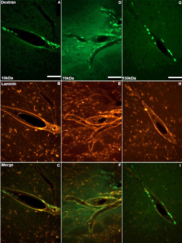
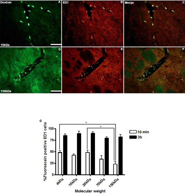
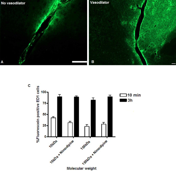
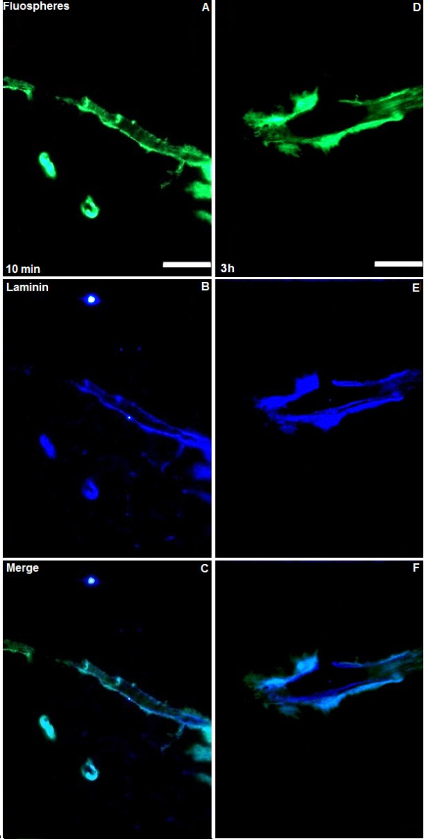
Methods: We analysed the perivascular distribution of 4, 10, 20, 70, 150 kDa fluorescein-labelled dextran and fluorescent nanoparticles at 10 min and 3 h following CED into rat striatum. We investigated the effect of local vasodilatation, slow infusion rates and ramping on the perivascular distribution of solutes. Co-localisation with perivascular basement membranes and vascular endothelial cells was identified by immunohistochemistry. The uptake of infusates by perivascular macrophages was quantified using stereological methods.

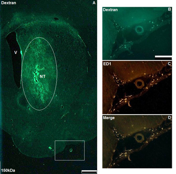
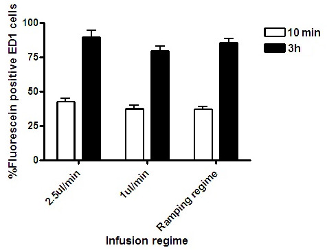
Results: Widespread perivascular distribution and macrophage uptake of fluorescein-labelled dextran was visible 10 min after cessation of CED irrespective of molecular weight. However, a significantly higher proportion of perivascular macrophages had taken up 4, 10 and 20 kDa fluorescein-labelled dextran than 150 kDa dextran (p < 0.05, ANOVA). Co-infusion with vasodilator, slow infusion rates and use of a ramping regime did not alter the perivascular distribution. CED of fluorescent nanoparticles indicated that particles co-localise with perivascular basement membranes throughout the striatum but, unlike soluble dextrans, are not taken up by perivascular macrophages after 3 h.

Conclusions: This study suggests that widespread perivascular distribution and interaction with perivascular macrophages is likely to be an inevitable consequence of CED of solutes. The potential consequences of perivascular distribution of therapeutic agents, and in particular cytotoxic chemotherapies, delivered by CED must be carefully considered to ensure safe and effective translation to clinical trials.


Figures

Figure 1
Figure 1
Widespread interstitial distribution of dextrans and co-localisation with leptomeningeal macrophages following CED. Figure A shows a low power micrograph of a coronal section through the right striatum of a rat prior to ED1 immunostaining (scale bar = 1 mm). Labelled are the right lateral ventricle (V), needle track (NT) and interstitial distribution of 150 kDa dextran at 3 h following CED (circled). High power micrographs of the area within the white box in Figure A illustrate co-localisation of 150 kDa dextran (green) with ED1-positive leptomeningeal macrophages (red) at 3 h (B, C and D, scale bar = 100 μm).
Figure 2
Figure 2
Co-localisation of solutes with basement membranes. Ten minutes after CED, 10, 70 and 150 kDa dextrans (green) were widely distributed throughout the perivascular basement membranes of the the striatum (A, D and G). Representative images of perivascular membranes immunostained for laminin (red) are shown in B, E and H. Figures C, F and I represent merged images. Scale bar = 100 μm.
Figure 3
Figure 3
Perivascular distribution of solutes external to vascular endothelium. Ten minutes after CED 10, 70 and 150 kDa dextrans (green) were seen in the perivascular spaces external to the vascular endothelium (A, D and G). Representative images of vascular endothelial cells immunostained for RECA1 (red) are shown in B, E and H. Figures C, F and I represent merged images. Scale bar = 100 μm.
Figure 4
Figure 4
Co-localisation of fluorescein-labelled dextran with ED1-positive perivascular macrophages. Ten minutes after CED there was widespread co-localisation of fluorescein-labelled dextran (green) with perivascular macrophages immunostained for ED1 (red). Representative images from animals infused with 10 and 150 kDa dextran are shown in A-F. Scale bar = 100 μm. Stereological cell counts demonstrated significantly higher co-localisation of 4, 10 and 20 kDa dextrans than 150 kDa dextran, 10 min after CED. For 4 and 20 kDa dextran, the increase in co-localisation was highly significant (*p < 0.01). However by 3 h there were no significant differences in uptake (figure G). Each histogram bar represents mean cell counts from 3 striata +/- S.D.
Figure 5
Figure 5
Co-infusion of 10 and 150 kDa dextran with nimodipine. Co-infusion of the vasodilator nimodipine did not prevent perivascular spread of solutes. Representative images at 10 min following CED of 150 kDa dextran (green) demonstrating widespread interstitial and perivascular solute are shown in figures A and B. Stereological comparisons of ED1 perivascular macrophage uptake of 10 and 150 kDa dextran with and without nimodipine did not reveal any statistically significant differences (figure C). Each histogram bar represents mean cell counts from 3 striata +/- S.D. Scale bars in both A and B = 100 μm.
Figure 6
Figure 6
CED using slow infusion rates and ramping regime. Slow infusion of 10 kDa dextran at 1 μl/min or use of a ramping regime did not prevent perivascular distribution. Stereological comparisons revealed no significant difference in uptake of 10 kDa Dextran by ED1-positive perivascular macrophages when infused at 1 or 2.5 μl/min or when a ramping regime was used. Each histogram bar represents mean cell counts from 3 striata +/- S.D.
Figure 7
Figure 7
CED of 20 nm fluorescent particles. Ten minutes following CED, 20 nm fluospheres (green, figure A) were seen to co-localise with perivascular basement membranes immunostained for laminin (blue, figure B). Appearances at 3 h following CED showed similar co-localisation without evidence of uptake of fluospheres by perivascular macrophages (figures D and E). Figures C and F represent merged images. Scale bar = 100 μm.

References

    1. Kroll RA, Pagel MA, Muldoon LL, Roman-Goldstein S, Neuwelt EA. Increasing volume of distribution to the brain with interstitial infusion: dose, rather than convection, might be the most important factor. Neurosurgery. 1996;38:746–752. doi: 10.1227/00006123-199604000-00024. discussion 752-744. - DOI - PubMed
    1. White E, Bienemann A, Malone J, Megraw L, Bunnun C, Wyatt M, Gill S. An evaluation of the relationships between catheter design and tissue mechanics in achieving high-flow convection-enhanced delivery. J Neurosci Methods. 2011;199:87–97. doi: 10.1016/j.jneumeth.2011.04.027. - DOI - PubMed
    1. Bruce JN, Fine RL, Canoll P, Yun J, Kennedy BC, Rosenfeld SS, Sands SA, Surapaneni K, Lai R, Yanes CL, Bagiella E, DeLaPaz RL. Regression of Recurrent Malignant Gliomas with Convection-Enhanced Delivery of Topotecan. Neurosurgery. 2011;69:1272–1279. doi: 10.1227/NEU.0b013e3182233e24. discussion 1279-1280. - DOI - PMC - PubMed
    1. Gill SS, Patel NK, Hotton GR, O'Sullivan K, McCarter R, Bunnage M, Brooks DJ, Svendsen CN, Heywood P. Direct brain infusion of glial cell line-derived neurotrophic factor in Parkinson disease. Nat Med. 2003;9:589–595. doi: 10.1038/nm850. - DOI - PubMed
    1. Bobo RH, Laske DW, Akbasak A, Morrison PF, Dedrick RL, Oldfield EH. Convection-enhanced delivery of macromolecules in the brain. Proc Natl Acad Sci USA. 1994;91:2076–2080. doi: 10.1073/pnas.91.6.2076. - DOI - PMC - PubMed

LinkOut - more resources