. 2012 Jan 19;11(1):81-90.
doi: 10.1016/j.chom.2011.11.012.

Mycobacterium tuberculosis inhibits neutrophil apoptosis, leading to delayed activation of naive CD4 T cells

Affiliations

Mycobacterium tuberculosis inhibits neutrophil apoptosis, leading to delayed activation of naive CD4 T cells

Robert Blomgran et al. Cell Host Microbe. .

Abstract

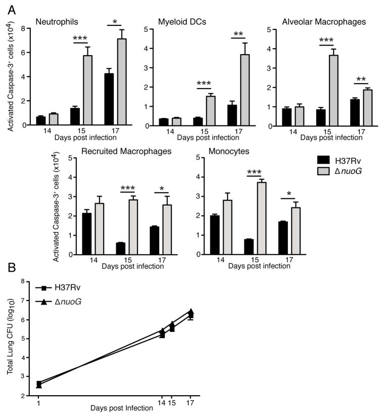
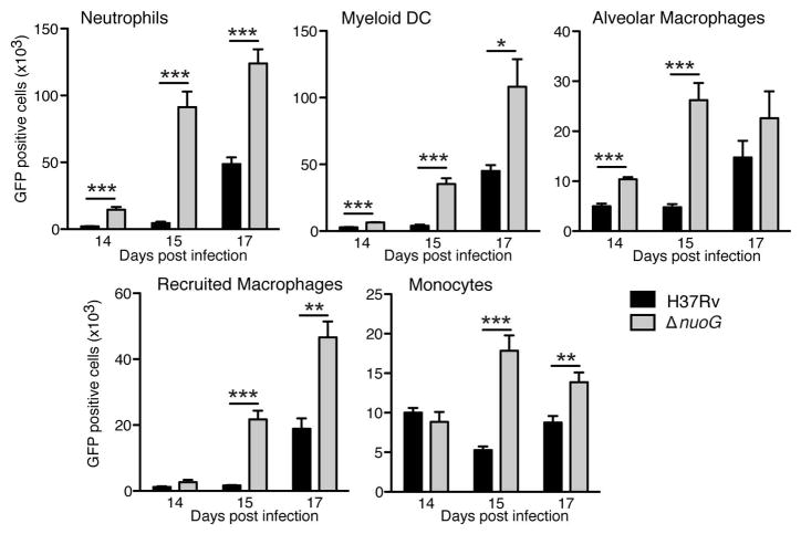
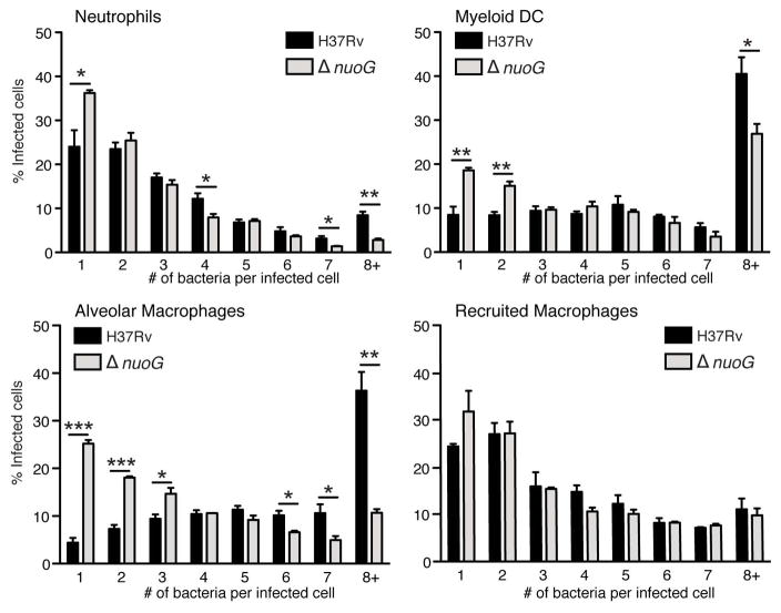
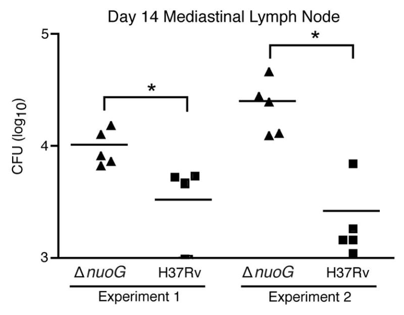
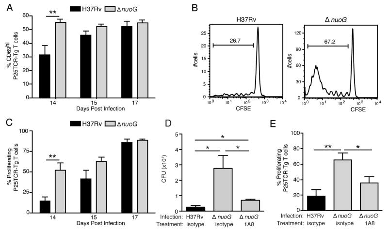
Mycobacterium tuberculosis promotes its replication by inhibiting the apoptosis of infected macrophages. A proapoptotic M. tuberculosis mutant lacking nuoG, a subunit of the type I NADH dehydrogenase complex, exhibits attenuated growth in vivo, indicating that this virulence mechanism is essential. We show that M. tuberculosis also suppresses neutrophil apoptosis. Compared to wild-type, the nuoG mutant spread to a larger number of lung phagocytic cells. Consistent with the shorter lifespan of infected neutrophils, infection with the nuoG mutant resulted in fewer bacteria per infected neutrophil, accelerated bacterial acquisition by dendritic cells, earlier trafficking of these dendritic cells to lymph nodes, and faster CD4 T cell priming. Neutrophil depletion abrogated accelerated CD4 T cell priming by the nuoG mutant, suggesting that inhibiting neutrophil apoptosis delays adaptive immunity in tuberculosis. Thus, pathogen modulation of apoptosis is beneficial at multiple levels, and enhancing phagocyte apoptosis promotes CD4 as well as CD8 T cell responses.


Figures

Figure 1
Figure 1
Increased frequency of apoptosis of diverse myeloid cells in lungs of mice infected with ΔnuoG M. tuberculosis. (A) Single cell preparations from lungs of C57BL/6J mice infected with wild-type H37Rv or ΔnuoG M. tuberculosis, were identified as neutrophils (Gr-1hi/CD11bhi), myeloid dendritic cells (CD11bhi/CD11chi), alveolar macrophages (CD11blow/CD11chi), recruited macrophages (CD11bhi/CD11cintermediate), or monocytes (CD11bhi/CD11cneg). After surface staining, fixation, and permeabilization, cells were stained for the presence of activated caspase-3. Cell numbers were calculated by multiplying the percentage in each subset obtained through flow cytometry by the total number of cells determined through manual count of the total number of cells from each mouse. (B) Growth of bacteria in the lungs after aerosol infection with the ΔnuoG and wild-type H37Rv strains of M. tuberculosis. There were no statistically significant differences in the number of bacteria of the two strains at any time point examined. Data in panel A–B are mean ± SD of five mice per group and time point, and each represent data from two separate experiments. *, p < 0.05; **, p < 0.01; ***, p < 0.001. See also Figure S1.
Figure 2
Figure 2
The proapoptotic ΔnuoG mutant is present in a larger number of diverse lung myeloid cells in vivo. C57BL/6J mice were infected with GFP-expressing wild-type H37Rv or ΔnuoG M. tuberculosis as in Figure 1, and the total number of infected cells in each myeloid cell subset was analyzed using flow cytometry detection of cells containing GFP-expressing bacteria. Data shown are mean ± SD of five mice per group and time point, and represent data from two separate experiments. *, p < 0.05; **, p < 0.01; ***, p < 0.001. See also Figure S2, Table S1.
Figure 3
Figure 3
The proapoptotic ΔnuoG mutant is present as fewer bacteria per infected cell than wild-type H37Rv. C57BL/6J mice were infected with GFP-expressing wild-type H37Rv or ΔnuoG M. tuberculosis, and GFP+ neutrophils, myeloid dendritic cells, alveolar macrophages, and recruited macrophages were sorted by FACS on day 17 post-infection. Lung cells from three mice were pooled prior to sorting, and three pools per group were analyzed, to ensure that 200–400 infected cells per pool could be examined by fluorescent microscopy. Cells containing 8 or more bacteria/cell are designated as 8+. Insufficient numbers of infected monocytes were obtained for analysis. Data are mean ± SD from 9 mice per group analyzed in pools of three mice per pool. *, p < 0.05; **, p < 0.01; ***, p < 0.001.
Figure 4
Figure 4
The proapoptotic ΔnuoG mutant traffics to the mediastinal lymph node earlier than wild-type H37Rv. C57BL/6 J mice were infected with wild-type H37Rv or ΔnuoG and the bacterial burden in the mediastinal lymph node was determined on the designated days postinfection. Data in experiment 1 are from the same experiment as displayed in Figures 1, 2, and 5(A–C). * designates p < 0.05.
Figure 5
Figure 5
The proapoptotic ΔnuoG mutant is associated with earlier activation of naïve M. tuberculosis Ag85B-specific CD4 T cells in vivo. Mice were infected one day after adoptive transfer of CFSE-labeled naïve P25TCR-Tg CD4+ T cells. (A) CD4 T cell activation as indicated by CD69 surface expression on P25TCR-Tg cells in the mediastinal lymph node on days 14, 15, and 17 post infection. (B) Representative CFSE dilution/cell proliferation profiles of adoptively-transferred P25TCR-Tg CD4+ cells in mediastinal lymph nodes of mice infected with H37Rv or ΔnuoG, 14 days post infection. The bars indicate the percentage of P25TCR-Tg cells that had undergone one or more cycles of proliferation. (C) Quantitation of of P25TCR-Tg CD4+ T cell proliferation in the mediastinal lymph nodes of groups of mice infected with H37Rv or ΔnuoG. (D) Neutrophil depletion abrogates the enhanced trafficking of M. tuberculosis in mice infected with ΔnuoG. Infected mice treated as in panels A and B received the Ly6G-specific neutrophil-depleting antibody 1A8 or isotype control antibody on day 8 post infection, and M. tuberculosis colony-forming units were quantitated on day 14 postinfection. (E) Neutrophil depletion abrogates the accelerated proliferation of Ag85B-specific CD4 T cells in mice infected with ΔnuoG. P25TCR-Tg CD4+ T cell proliferation in the mediastinal lymph nodes of the same mice as in panel D. Data in panels A–E are mean ± SD of five mice per group and time point, where A–C is representative of three separate experiments, and D and E of two separate and independent experiments. *, p < 0.05; **, p < 0.01.

References

    1. Abadie V, Badell E, Douillard P, Ensergueix D, Leenen PJ, Tanguy M, Fiette L, Saeland S, Gicquel B, Winter N. Neutrophils rapidly migrate via lymphatics after Mycobacterium bovis BCG intradermal vaccination and shuttle live bacilli to the draining lymph nodes. Blood. 2005;106:1843–1850. - PubMed
    1. Balcewicz-Sablinska MK, Keane J, Kornfeld H, Remold HG. Pathogenic Mycobacterium tuberculosis evades apoptosis of host macrophages by release of TNF-R2, resulting in inactivation of TNF-alpha. J Immunol. 1998;161:2636–2641. - PubMed
    1. Behar SM, Divangahi M, Remold HG. Evasion of innate immunity by Mycobacterium tuberculosis: is death an exit strategy? Nat Rev Microbiol. 2010;8:668–674. - PMC - PubMed
    1. Berry MP, Graham CM, McNab FW, Xu Z, Bloch SA, Oni T, Wilkinson KA, Banchereau R, Skinner J, Wilkinson RJ, et al. An interferon-inducible neutrophil-driven blood transcriptional signature in human tuberculosis. Nature. 2010;466:973–977. - PMC - PubMed
    1. Blomgran R, Ernst JD. Lung neutrophils facilitate activation of naive antigen-specific CD4+ T cells during Mycobacterium tuberculosis infection. J Immunol. 2011;186:7110–7119. - PMC - PubMed

Publication types

MeSH terms