. 2012 Jan 17;22(1):116-30.
doi: 10.1016/j.devcel.2011.10.030.

Phosphoinositide signaling regulates the exocyst complex and polarized integrin trafficking in directionally migrating cells

Affiliations

Phosphoinositide signaling regulates the exocyst complex and polarized integrin trafficking in directionally migrating cells

Narendra Thapa et al. Dev Cell. .

Abstract

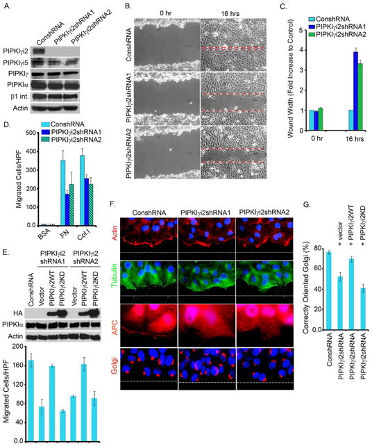
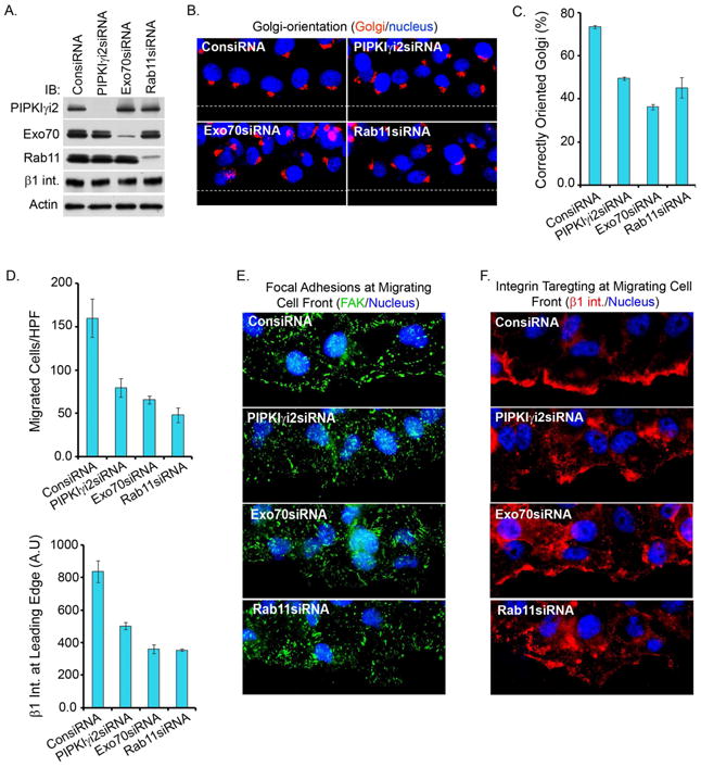
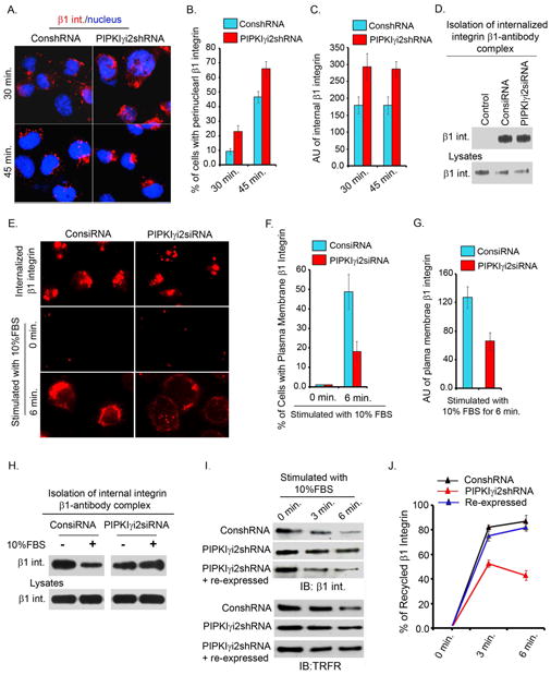
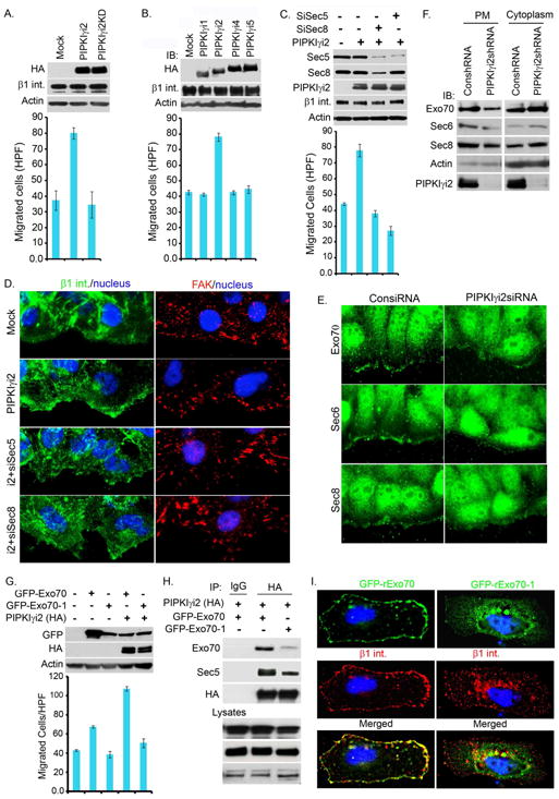
Polarized delivery of signaling and adhesion molecules to the leading edge is required for directional migration of cells. Here, we describe a role for the PIP(2)-synthesizing enzyme, PIPKIγi2, in regulation of exocyst complex control of cell polarity and polarized integrin trafficking during migration. Loss of PIPKIγi2 impaired directional migration, formation of cell polarity, and integrin trafficking to the leading edge. Upon initiation of directional migration, PIPKIγi2 via PIP(2) generation controls the integration of the exocyst complex into an integrin-containing trafficking compartment that requires the talin-binding ability of PIPKIγi2, and talin for integrin recruitment to the leading edge. A PIP(2) requirement is further emphasized by inhibition of PIPKIγi2-regulated directional migration by an Exo70 mutant deficient in PIP(2) binding. These results reveal how phosphoinositide generation orchestrates polarized trafficking of integrin in coordination with talin that links integrins to the actin cytoskeleton, processes that are required for directional migration.


Figures

Figure 1
Figure 1. PIPKIγi2 is Required for Directional Cell Migration
(A) shRNA/lentiviral system was used to knockdown endogenous PIPKIγi2. Isolated cell lines (designated as PIPKIγi2shRNA1 and PIPKIγi2shRNA2) were examined by immunoblotting for knockdown of PIPKIγi2. (B) Control or PIPKIγi2 knockdown cells grown to confluency were wounded and wound width measured at zero and 16 hours post-wounding (representative images at zero and 16 hours post-wounding). (C) The results are expressed as average fold increase in wound width compared with control cells at zero and 16 hours post-wounding (mean±SD from three independent experiments). (D) For haptotactic cell migration towards FN or Col.I, the modified Boyden chamber was used. Results expressed as the total number of cells migrated/HPF (mean ±SD from three independent experiments). (E) Rescue of cell migration defect in PIPKIγi2 knockdown cells. Lentiviral expression system was used to express PIPKIγi2 or its kinase dead mutant into the PIPKIγi2 knockdown cells. Haptotactic cell migration was examined as described above (results are mean ±SD from three independent experiments) (F) Confluent cell cultures were wounded and processed 6-hours post-wounding to examine orientation of actin (red), microtubules (green), APC (red) and Golgi (red) towards the direction of cell migration. (G) Quantitative data for Golgi orientation (mean±SD of three independent experiments). See also Figure S1, Movie S1 and S2
Figure 2
Figure 2. Knockdown of PIPKIγi2, Exocyst Complex components or Rab11 Impairs Polarized Recruitment of β-integrins and Cell Migration
(A) Knockdown of PIPKIγi2, Exo70 or Rab11 in MDA-MB-231 cells. Cells were transfectd with specific siRNAs and knockdown of indicated proteins was examined by immunobloting. (B) Confluent cell culture (48-72 hours post-transfection with siRNA), were wounded and processed 6 hours post-wounding to examine Golgi (red) orientation towards the direction of cell migration. (C) Quantitative data of Golgi orientation (mean±SD of three independent experiments). (D) The modified Boyden chamber assay as described above was used to examine the migration of siRNA-treated cells towards FN. Results were expressed as the total number of cells migrated/HPF (mean±SD from three independent experiments). (E, F) Confluent cell culture (48-72 hours post-transfection with siRNA) were fixed 5-6 hours post-wounding and immunostained for FAK (green) and β1-integrin (red) to examine the focal adhesion complex formation and β1-integrin recruitment at the leading edge. (G) Quantitative data of β1 integrin recruitment at the migrating cell fronts. The average fluorescence intensity (AU) of β1 integrin at migrating cell front was measured using the metamorph (mean±SD of three independent experiments). See also Figure S2
Figure 3
Figure 3. Cell Migration Promotes PIPKIβi2 Reorganization and Association with β1-integrin Complexes
(A) PIPKIγi2 colocalizes with β1-integrins and talin. MDA-MB-231 cells expressing moderate level of HA-tagged PIPKIγi2 were wounded and processed for immunofluorescence (4-5 hours post-scratching). PIPKIγi2 (red) and β1-integrin or talin (green) are recruited to and colocalize at migrating cell fronts and at intracellular compartments. (B) PIPKIγi2 colocalizes with Rab4 and Rab11. MDA-MB-231 cells expressing PIPKIγi2 were seeded on FN-coated coverslips and cultured for 2-3 hours prior to cell fixation and immunostaining for PIPKIγi2 (green) and Rab4 or Rab11 (red). (C) Cell migration enhances a PIPKIγi2 association with β1-integrin and talin. Confluent MDA-MB-231 or HeLa cells were wounded extensively so that about 50% of cells were detached from culture plates. Cells were harvested at different time points and β1-integrin was immunoprecipitated followed by immunoblotting to examine the co-immunoprecipitation of PIPKIγi2 and talin. (P.S., post-scratch). (D) PIPKIγi2 is required for talin association with β1-integrin in migrating Cells. Confluent cultures of control or PIPKIγi2 knockdown cells (HeLa) were wounded as described above before immunoprecipitation of β1-integrin to examine the co-immunoprecipitation of talin and PIPKiγi2 by immunoblotting. (E) Cytoplasmic domain of β1-integrin pulled down both talin and PIPKIγi2. GST-fusion protein of cytoplasmic domain of β1- or α5-integrin was incubated with cell lystates prepared from MDA-MB-231 cells expressing PIPKIγ1, PIPKIγi2 or PIPKIγi2Y649F. Pull down of talin and PIPKIγi2 were examined by immunoblotting. (F) In vitro binding study. GST-fusion protein of cytoplasmic domain of β1- or α5-integrin was incubated with His-tagged PIPKIγi2 purified from bacteria and PIPKIγi2 binding examined by immunoblotting using anti-His antibody. See also Figure S3
Figure 4
Figure 4. PIPKIγi2 Knockdown Impairs β1-integrin Exocytosis
(A) For β1-integrin endocytosis, cell surface β1-integrin were labeled with anti-β1 antibody at 4°C. Cells were incubated at 37°C to induce internalization. Shown is the β1-integrin internalized after 30 and 45 minutes incubation. (B) Cells with distinct perinuclear accumulation of β1-integrin-antibody complex were counted and expressed as % of total cells. A total of 150-200 cells were counted for each condition (results are mean±SD of three independent experiments). (C) Average fluorescence intensity (AU) of internalized β1-integrin in knockdown and control cells was measured (around 150 cells included for each condition; results are mean±SD of three independent experiments). (D) For biochemical assay of β1-integrin endocytosis, cell surface β1-integrin were labeled with anti-β1 antibody at 4°C followed by incubation of cells at 37°C for 10 minutes to induce internalization. The content of internalized β1-integrins in control or PIPKIγi2 knockdown cells was examined by immunoblotting. (E) For examining β1-integrin accumulation at perinuclear regions, cells were permeabilized (top panel) before immunostaining as described in “Experimental Procedures”. Cells were processed for immunostaining without cell permeabilization to examine the β1-integrin (red) trafficking to the plasma membrane before (middle panels) or after (bottom panels) cell stimulation with FBS. (F) The number of cells with distinct plasma-membrane localization of β1-integrin in control vs. PIPKIγi2 knockdown cells was quantified (around 150 cells counted each time; values are mean±SD of three independent experiments). (G) Average fluorescence intensity (AU) of plasma-membrane localization of β1-integrin in control vs. PIPKIγi2 knockdown cells (around 150 cells counted each time; values are mean±SD of three independent experiments). (H) The content of internal β1-integrin after FBS stimulation. Representative image of three independent experiments showing that PIPKIγi2 knockdown slowed β1-integrin trafficking to the plasma membrane. (I) Integrin recycling was examined by cell surface biotinylation assay as described in “Experimenal Procedures”. Biotinylated cell surface proteins remaining inside the cells were isolated using streptavidine affinity gel followed by examination of β1-integrin and transferine receptor (TRFR) by immunoblotting. (J) Quantitative data of β1-integrin recycling. The % of β1-integrin recycled was calculated as described in “Experimental Procedures” (values are mean±SD from three independent experiments).
Figure 5
Figure 5. PIPKIγi2 Directly Associates with the Exocyst Complex
(A) Endogenous PIPKIγi2 was immunoprecipitated from MDA-MB-231 cells and co-imunoprecipitation of exocyst complex examined by immunoblotting. (B) MDA-MB-231 cells expressing moderate level of HA-tagged PIPKIγi2 grown to confluence were harvested at different time points after scratch-wounding. PIPKIγi2 was immunoprecipitated using anti-HA antibody and co-immunoprecipitation of β1-integrin and exocyst complex examined by immunoblotting. (C) HeLa cells grown to confluence were harvested at different time points after scratch-wounding. β1-integrin was immunoprecipitated and co-immunoprecipitation of PIPKIγi2 and exocyst components examined by immunoblotting. (D) MDA-MB-231 cells expressing PIPKIγi1, PIPKIγi2, PIPKIγi2KD or PIPKIγi2Y649F were harvested 2-3 hours post-wounding. β1-integrin was immunoprecipitated and co-immunoprecipitation of exocyst complex proteins examined by immunoblotting. (E) MDA-MB-231 cells expressing PIPKIγi1, PIPKIγi2, PIPKIγi2KD or PIPKIγi2Y649F were harvested 2-3 hours post-wounding. PIPKIγi2 and other mutants were immunoprecipitated using anti-HA antibody and co-immunoprecipitation of exocyst complex examined by immunoblotting. (F) Sec6 and Exo70 directly interact with PIPKIγi2. GST-fusion protein of exocyst complex components were incubated with His-PIPKIγi2 purified from bacteria. PIPKIγi2 binding was examined by immunoblotting using anti-His antibody. (G) Exo70 and Sec6 co-immunoprecipitate endogenous PIPKIγi2. HeLa cells were transiently transfected with Flag-tagged Exo70 or Sec6 and immunoprecipitated using anti-Flag antibody. Co-immunoprecipitation of PIPKIγi2 and other components of exocyst complex were examined by immunoblotting. (H) Knockdown of Exo70 or Sec6 impairs PIPKIγi2 association with exocyst complex. HeLa cells were transfected with siRNA for Exo70 or Sec6. 24 hours after the siRNA transfection, cells were transfected with HA-tagged PIPKIγi2. Next day, cells were harvested to immunoprecipitate PIPKIγi2 and co-immunoprecipitation of exocyst complex examined by immunoblotting. See also Figure S4
Figure 6
Figure 6. The Exocyst Complex is Required for PIPKIγi2-regulated Cell Migration
(A) PIPKIγi2 expression promotes cell migration. HeLa cells transiently transfected with PIPKIγi2 or PIPKIγi2KD were monitored for changes in haptotactic cell migration using a modified Boyden chamber assay. The results are expressed as migrated cells/HPF (mean±SD of three experiments). Immunoblots were used to examine PIPKIγ expression using anti-HA antibody. (B) Cell migration assays were performed as above in HeLa cells transiently transfected with PIPKIγ isoforms and results expressed as migrated cells/HPF (mean±SD of three experiments). Immunoblots were used to examine PIPKIγ expression using anti-HA antibody. (C) Cell migration assays were performed in HeLa cells treated with siRNA to knockdown exocyst components (Sec5 or Sec8) followed by PIPKIγi2 overexpression as described above. Results expressed as migrated cells/HPF (mean±SD of three experiments). Knockdown of Sec5 or Sec8 and expression of PIPKIγi2 were monitored by immunoblotting. (D) Exocyst complex is required for polarized recruitment of β1-integrin. HeLa cells stably expressing PIPKIγi2 were treated with siRNA to knockdown Sec5 or Sec8. 48-72 hours post-transfection, cells were scratch-wounded and immunostained for β1-integrin (green) and FAK (red) to examine the recruitment of β1-integrin and focal adhesion formation at migrating cell fronts. (E) Confluent culture of cells (48-72 hours post-transfection with siRNA) were processed 2-3 hours post-wounding. The polarized recruitment of endogenous exocyst complex (Exo70, Sec6 and Sec8) (green) to migrating cell fronts were examined using their specific antibodies. (F) Crude plasma membrane was isolated from control or PIPKIγi2 knockdown cells followed by examination of exocyst complex components in plasma membrane and cytosol. (G) HeLa cells were transfected with GFP-Exo70 or GFP-Exo70-1 or cotransfected with PIPKIγi2. Haptotactic cell migration towards FN was examined as described above. The results expressed as migrated cells/HPF (mean±SD of three experiments). (H) HeLa cells were transfected with either GFP-Exo70 or GFP-Exo70-1. GFP-Exo70 colocalized with β1-integrin (red) at plasma membrane whereas GFP-Exo70-1 was found either diffusely distributed into the cytoplasma or accumulated around perinuclear regions. β1-integrin was poorly recruited to plasma membrane and accumulated around perinuclear regions in GFP-Exo70-1 expressing cells. (I) Exo70-1 poorly associates with PIPKIγi2 and impairs the PIPKIγi2 association with the exocyst complex. HeLa cells were cotransfected with PIPKIγi2 and Exo70 or Exo70-1. Cells were harvested 24 hours post-transfection to immunoprecipitate PIPKIγi2 and co-immunoprecipitation of exocyst complex examined by immunoblotting. See also Figure S5
Figure 7
Figure 7. PIPKIγi2 Integrates Exocyst Complex and Talin with Integrin, and is Required for Integrin Trafficking
(A) HeLa cells cotransfected with Flag-tagged exocyst complex components (Sec6 or Sec8 or Exo70) and HA-tagged PIPKIγi2 were allowed to adhere on FN-coated coverslips for 1-2 hours before immunostaining with anti-Flag (green) and anti-HA (red) antibodies. (B) PIPKIγi2, Exo70 and α5-GFP integrin colocalize at cell membrane and intracellular compartments. HeLa cells (bottom) or HeLa cells stably expressing HA-tagged PIPKIγi2 (top) were co-transfected with Flag-tagged Exo70 and α5 integrin-GFP or GFP. Cells were fixed and immunostained using anti-HA (blue) or anti-Flag (red) antibodies. (C) Exo70 colocalizes with talin at focal adhesions and intracellular sites. HeLa cells transiently transfected with Flag-tagged Exo70 alone or cotransfected with HA-tagged PIPKIγi2 were allowed to adhere on FN-coated coverslips for 1-2 hours before immunostaining with anti-HA (blue), anti-Flag (red) and anti-talin (green) antibodies. (D) PIPKIγi2 integrates talin, β1-integrin and exocyst complex in the same complex. HeLa cells were transfected with PIPKIγi1 or PIPKIγi2. Talin were immunoprecipitated 24 hours post-transfection followed by immunoblotting for exocyst complex and β1-integrin. (E) PIPKIγi2 were immunoprecipitated from HeLa cells stably expressing PIPKIγi2 or PIPKIγi2KD at different time points following wounding to induce migration. Immunocomplexes were examined for presence of integrins and/or talin by immunoblotting. (F) Model depicting the role of PIPKIγi2 in integrin trafficking in directionally migrating cells. Cell migration induces the integration of PIPKIγi2, talin, β1-integrin into the complex either in plasma membrane or in intracellular recycling compartments. Further, PIP2 generation by PIPKIγi2 into the complex facilitates the assembly of the exocyst complex. Thus, coordinated activity of PIPKIγi2 and the exocyst complex in concert with talin promotes the polarized recruitment and trafficking of integrin molecules to migrating cell fronts. Loss of PIPKIγi2 or the exocyst complex or talin compromises the polarized recruitment/trafficking of integrin impairing cell polarization and directional cell migration. See also Figure S6

References

    1. Anderson RA, Boronenkov IV, Doughman SD, Kunz J, Loijens JC. Phosphatidylinositol phosphate kinases, a multifaceted family of signaling enzymes. J Biol Chem. 1999;274:9907–9910. - PubMed
    1. Bairstow SF, Ling K, Su X, Firestone AJ, Carbonara C, Anderson RA. Type Igamma661 phosphatidylinositol phosphate kinase directly interacts with AP2 and regulates endocytosis. J Biol Chem. 2006;281:20632–20642. - PubMed
    1. Bretscher MS. Endocytosis: relation to capping and cell locomotion. Science. 1984;224:681–686. - PubMed
    1. Bretscher MS. Endocytosis and recycling of the fibronectin receptor in CHO cells. EMBO J. 1989;8:1341–1348. - PMC - PubMed
    1. Caswell P, Norman J. Endocytic transport of integrins during cell migration and invasion. Trends Cell Biol. 2008;18:257–263. - PubMed

Publication types

MeSH terms