Multicenter Study
. 2012 Feb;153(2):204-208.e1.
doi: 10.1016/j.ajo.2011.11.035.

An outbreak of streptococcus endophthalmitis after intravitreal injection of bevacizumab

Affiliations
Multicenter Study

An outbreak of streptococcus endophthalmitis after intravitreal injection of bevacizumab

Roger A Goldberg et al. Am J Ophthalmol. 2012 Feb.

Abstract

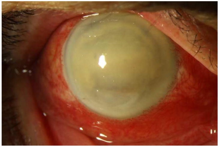
Purpose: To report a series of patients with Streptococcus endophthalmitis after injection with intravitreal bevacizumab prepared by the same compounding pharmacy.

Design: Noncomparative consecutive case series.

Methods: Medical records and microbiology results of patients who presented with endophthalmitis after injection with intravitreal bevacizumab between July 5 and July 8, 2011, were reviewed.

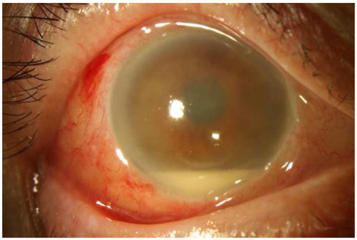
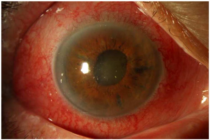
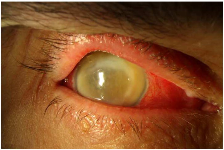
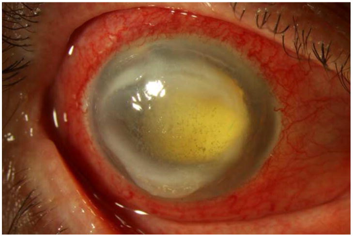
Results: Twelve patients were identified with endophthalmitis, presenting 1 to 6 days after receiving an intravitreal injection of bevacizumab. The injections occurred at 4 different locations in south Florida. All patients received bevacizumab prepared by the same compounding pharmacy. None of the infections originated at the Bascom Palmer Eye Institute, Miami, Florida, although 9 patients presented to its tertiary-care ophthalmic emergency room for treatment, and 3 additional patients were seen in consultation. All patients were treated initially with a vitreous tap and injection; 8 patients subsequently received a vitrectomy. Microbiology cultures for 10 patients were positive for Streptococcus mitis/oralis. Seven unused syringes of bevacizumab prepared by the compounding pharmacy at the same time as those prepared for the affected patients also were positive for S. mitis/oralis. After 4 months of follow-up, all but 1 patient had count fingers or worse visual acuity, and 3 required evisceration or enucleation. Local, state, and federal health department officials have been investigating the source of the contamination.

Conclusions: In this outbreak of endophthalmitis after intravitreal bevacizumab injection, Streptococcus mitis/oralis was cultured from the majority of patients and from all unused syringes. Visual outcomes were generally poor. The most likely cause of this outbreak was contamination during syringe preparation by the compounding pharmacy.


Figures

Figure 1
Figure 1. Streptococcus mitis/oralis endophthalmitis case example – improving clinical picture
Case 2 presented 2 days after intravitreal bevacizumab injection with hand motions vision (left). One week after initial vitreous tap and inject (middle), the hypopyon showed marked contraction, though the vision was light perception. By one month (right), the clinical appearance had improved, but the vision was still light perception. After subsequent vitrectomy, the visual acuity was hand motions.
Figure 2
Figure 2. Streptococcus mitis/oralis endophthalmitis case example – prominent ring infiltrate
Case 8 presented 6 days after intravitreal bevacizumab injection with light perception vision (left). One week after initial vitreous tap and inject (middle), both the corneal ring infiltrate and the anterior chamber fibrin showed signs of contracting. By one month (right), the ring infiltrate had improved and the anterior chamber was nearly clear, though visual acuity was still light perception.
Figure 3
Figure 3. Streptococcus mitis/oralis endophthalmitis case example – eviscerated six weeks after injection
Case 9 presented 6 days after intravitreal bevacizumab injection with light perception vision (left). One week after initial vitreous tap and inject (middle) the vision had deteriorated to no light perception. At one month (right), the visual acuity was still no light perception; ultimately the patient underwent an evisceration at six weeks after initial presentation.

References

    1. McCannel CA. Meta-analysis of endophthalmitis following intravitreal injection of anti-VEGF agents: causative organisms and possible prevention strategies. Retina. 2011;31(4):654–661. - PubMed
    1. Moshfeghi AA, Rosenfeld PJ, Flynn HW, Jr, et al. Endophthalmitis after intravitreal anti-vascular endothelial growth factor antagonists: a six-year experience at a university referral center. Retina. 2011;31(4):662–668. - PubMed
    1. The CATT Research Group. Ranibizumab and bevacizumab for neovascular age-related macular degeneration. N Engl J Med. 2011;364(20):1897–1908. - PMC - PubMed
    1. Brechner RJ, Rosenfeld PJ, Babish JD, Caplan S. Pharmacotherapy for Neovascular Age-Related Macular Degeneration: An Analysis of the 100% 2008 Medicare Fee-For-Service Part B Claims File. Am J Ophthalmol. 2011;151(5):887–895. - PubMed
    1. Miller JM, Scott IU, Flynn HW, Jr, Smiddy WE, Corey RP, Miller D. Endophthalmitis caused by Streptococcus pneumoniae. Am J Ophthalmol. 2004;138(2):231–236. - PubMed

Publication types

MeSH terms