Diagnostic capability of spectral-domain optical coherence tomography for glaucoma
- PMID: 22265147
- PMCID: PMC3517739
- DOI: 10.1016/j.ajo.2011.09.032
Diagnostic capability of spectral-domain optical coherence tomography for glaucoma
Abstract
Purpose: To determine the diagnostic capability of spectral-domain optical coherence tomography in glaucoma patients with visual field defects.
Design: Prospective, cross-sectional study.
Settings: Participants were recruited from a university hospital clinic.
Study population: One eye of 85 normal subjects and 61 glaucoma patients with average visual field mean deviation of -9.61 ± 8.76 dB was selected randomly for the study. A subgroup of the glaucoma patients with early visual field defects was calculated separately.
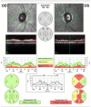
Observation procedures: Spectralis optical coherence tomography (Heidelberg Engineering, Inc) circular scans were performed to obtain peripapillary retinal nerve fiber layer (RNFL) thicknesses. The RNFL diagnostic parameters based on the normative database were used alone or in combination for identifying glaucomatous RNFL thinning.
Main outcome measures: To evaluate diagnostic performance, calculations included areas under the receiver operating characteristic curve, sensitivity, specificity, positive predictive value, negative predictive value, positive likelihood ratio, and negative likelihood ratio.
Results: Overall RNFL thickness had the highest area under the receiver operating characteristic curve values: 0.952 for all patients and 0.895 for the early glaucoma subgroup. For all patients, the highest sensitivity (98.4%; 95% confidence interval, 96.3% to 100%) was achieved by using 2 criteria: ≥ 1 RNFL sectors being abnormal at the < 5% level and overall classification of borderline or outside normal limits, with specificities of 88.9% (95% confidence interval, 84.0% to 94.0%) and 87.1% (95% confidence interval, 81.6% to 92.5%), respectively, for these 2 criteria.
Conclusions: Statistical parameters for evaluating the diagnostic performance of the Spectralis spectral-domain optical coherence tomography were good for early perimetric glaucoma and were excellent for moderately advanced perimetric glaucoma.
Copyright © 2012 Elsevier Inc. All rights reserved.
Figures

References
-
- Wollstein G, Beaton S, Paunescu A, et al. Optical coherence tomography in glaucoma. In: Schuman JS, Puliafito CA, Fujimoto JG, editors. Optical coherence tomography of ocular diseases. SLACK Incorporated; Thorofare, NJ: 2004. pp. 483–610.
-
- Blumenthal EZ, Weinreb RN. Assessment of the retinal nerve fiber layer in clinical trials of glaucoma neuroprotection. Surv Ophthalmol. 2001;45(Suppl 3):S305–12. discussion S332-4. - PubMed
-
- Quigley HA, Addicks EM, Green WR. Optic nerve damage in human glaucoma. III. Quantitative correlation of nerve fiber loss and visual field defect in glaucoma, ischemic neuropathy, papilledema, and toxic neuropathy. Arch Ophthalmol. 1982;100(1):135–46. - PubMed
-
- Sommer A, Katz J, Quigley HA, et al. Clinically detectable nerve fiber atrophy precedes the onset of glaucomatous field loss. Arch Ophthalmol. 1991;109(1):77–83. - PubMed
Publication types
MeSH terms
Grants and funding
LinkOut - more resources
Full Text Sources
Medical
Miscellaneous

