

Comment
doi: 10.1016/j.cell.2011.12.020.
RIP3 finds partners in crime
Affiliations
- PMID: 22265396
- PMCID: PMC3571655
- DOI: 10.1016/j.cell.2011.12.020
Item in Clipboard
Comment
RIP3 finds partners in crime
Cell.
.
Abstract
Programmed necrosis has long been recognized as a crucial component of animal development; however, the signaling pathway beyond the protein kinases RIP1 and RIP3 remains largely unknown. Sun et al. and Wang et al. now identify critical factors downstream of RIP1 and RIP3 in programmed necrosis, extending our understanding of this form of cell death.
Copyright © 2012 Elsevier Inc. All rights reserved.
Figures
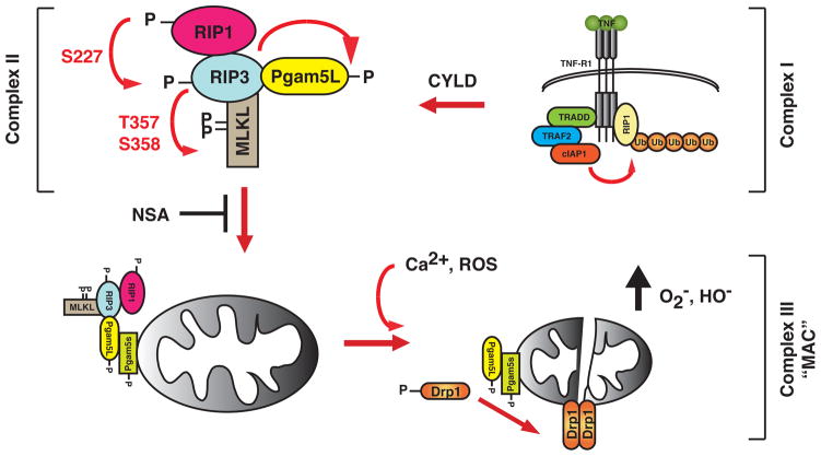
Sequential recruitment and activation of necrosis signaling complexes. RIP1 is recruited to the activated TNFR-1 undergoes heavy ubiquitination by E3 ligases such as TRAF2 and cIAP-1. This membrane and receptor associated complex, termed Complex I, is responsible for NF-κB activation. As the membrane-associated Complex I become internalized, deubiquitinases such as cylindromatosis (CYLD) removes the polyubiquitin chains on RIP1. The deubiquitination of RIP1 and inhibition of caspase 8 is crucial for the assembly of the secondary signaling complex (Complex II). At this cytoplasmic complex, RIP1 likely phosphorylates RIP3 at S227, which in turn phosphorylates PGAM5L and MLKL at T357 and S358. These phosphorylation events are important for the RIP3 necrosis signaling complex to engage PGAM5s on the mitochondrial membrane, a step that is inhibited by the small molecule inhibitor NSA. Once activated by phosphorylation, the PGAM5L/PGAM5s complex dephosphorylates the mitochondrial fission regulator Drp1 to induce its dimerization and activation. Excessive Drp1 activity could lead to disruption of mitochondrial functions and other organelle and membrane damages that cumulates in programmed necrosis. The PGAM5L-PGAM5s-Drp1 mitochondrial attack complex (MAC) could also be activated by calcium flux and surge of intracellular reactive oxygen species (ROS).
Comment on
-
Mixed lineage kinase domain-like protein mediates necrosis signaling downstream of RIP3 kinase.Cell. 2012 Jan 20;148(1-2):213-27. doi: 10.1016/j.cell.2011.11.031. Cell. 2012. PMID: 22265413
-
The mitochondrial phosphatase PGAM5 functions at the convergence point of multiple necrotic death pathways.Cell. 2012 Jan 20;148(1-2):228-43. doi: 10.1016/j.cell.2011.11.030. Cell. 2012. PMID: 22265414
References
-
- He SD, Wang L, Miao L, Wang T, Du FH, Zhao LP, Wang XD. Receptor Interacting Protein Kinase-3 Determines Cellular Necrotic Response to TNF-alpha. Cell. 2009;137:1100–1111. - PubMed
Publication types
Grants and funding
LinkOut - more resources
Full Text Sources
Miscellaneous
