. 2012 Jan 20;148(1-2):72-83.
doi: 10.1016/j.cell.2011.11.026.

Foxa1 and Foxa2 are essential for sexual dimorphism in liver cancer

Affiliations

Foxa1 and Foxa2 are essential for sexual dimorphism in liver cancer

Zhaoyu Li et al. Cell. .

Abstract

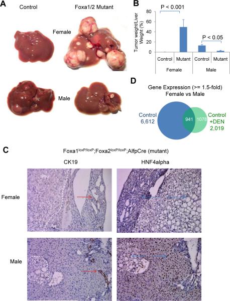
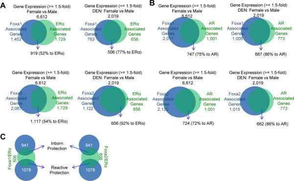
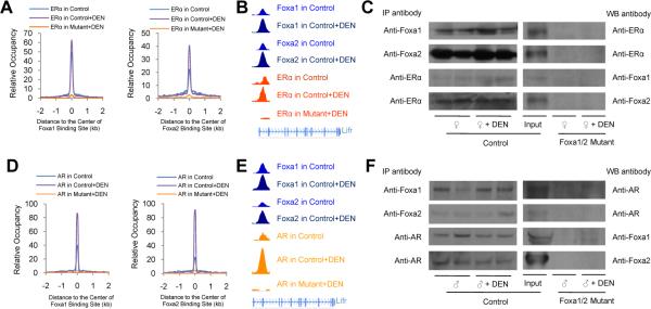
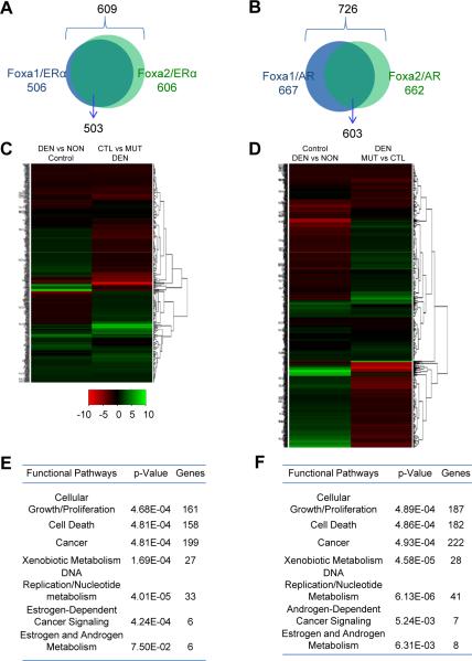
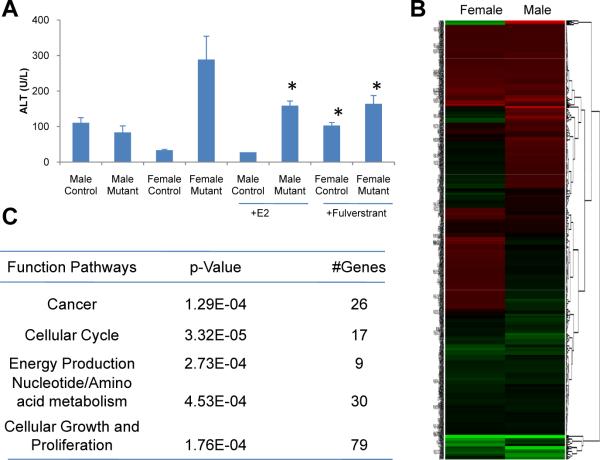
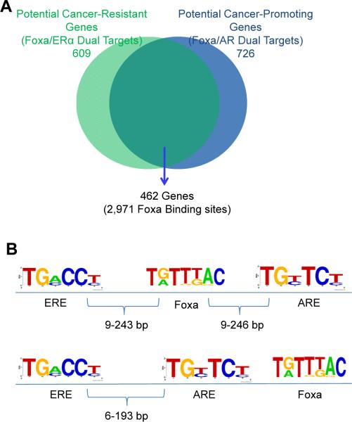
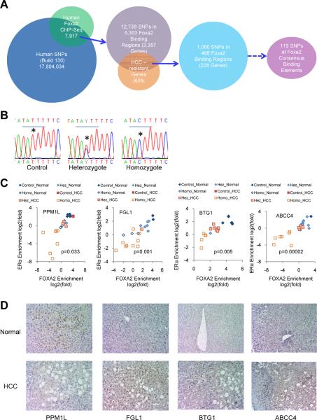
Hepatocellular carcinoma (HCC) is sexually dimorphic in both rodents and humans, with significantly higher incidence in males, an effect that is dependent on sex hormones. The molecular mechanisms by which estrogens prevent and androgens promote liver cancer remain unclear. Here, we discover that sexually dimorphic HCC is completely reversed in Foxa1- and Foxa2-deficient mice after diethylnitrosamine-induced hepatocarcinogenesis. Coregulation of target genes by Foxa1/a2 and either the estrogen receptor (ERα) or the androgen receptor (AR) was increased during hepatocarcinogenesis in normal female or male mice, respectively, but was lost in Foxa1/2-deficient mice. Thus, both estrogen-dependent resistance to and androgen-mediated facilitation of HCC depend on Foxa1/2. Strikingly, single nucleotide polymorphisms at FOXA2 binding sites reduce binding of both FOXA2 and ERα to their targets in human liver and correlate with HCC development in women. Thus, Foxa factors and their targets are central for the sexual dimorphism of HCC.


Figures

Figure 1
Figure 1. Foxa1 and Foxa2 Protect Female Mice from HCC and Promote HCC in Male Mice
(A) Livers from Foxa1loxP/loxP;Foxa2loxP/loxP (control) and Foxa1loxP/loxP;Foxa2loxP/loxP;AlfpCre (mutant) mice 18 weeks after exposure to hepatic carcinogens. (B) The percentage of tumor weight to liver weight assayed by NMR. 5–6 mice were analyzed in each group. (C) Immunostaining of cytokeratin 19 (CK19) and hepatic nuclear factor 4 alpha (HNF4alpha) in livers from Foxa1loxP/loxP;Foxa2loxP/loxP;AlfpCre mice. Tumors are delimited by dotted white lines. Arrows indicate positive staining for CK19 (red) and HNF4alpha (blue). (D) Gender-differential gene expression in control mice with or without carcinogen (DEN) administration. Purified hepatic RNA from four groups of mice (female and male, exposed to DEN or not) were processed for expression profiling. Over 6,000 genes are expressed in gender-specific fashion in control mice, but only ~ 2,000 in mice exposed to carcinogen. See also Figure S1.
Figure 2
Figure 2. Enhanced Co-Regulation of Foxa and ERα/AR during Hepatocarcinogenesis
Livers were collected from Foxa1loxP/loxP;Foxa2loxP/loxP (control) and Foxa1loxP/loxP;Foxa2loxP/loxP;AlfpCre (mutant) mice with/without carcinogen exposure for 18 weeks and processed for ChIP-Seq analysis. (A, B) Genome-wide co-regulation by Foxa1/2 and ERα (A) or AR (B) in control liver with or without carcinogen treatment. Genes associated with Foxa1/2, ERα, or AR were identified from ChIP-Seq analyses. (C) Foxa/ERα dual-target genes potentially involved in resistance to hepatocellular carcinoma (HCC) in the liver of female mice before and after carcinogen administration, defined as inborn and reactive protection, respectively. See also Figure S2, and Table S1 and S2.
Figure 3
Figure 3. Both Estrogen Signaling Preventing HCC and Androgen Signaling Promoting HCC Depend on Foxa1/2
Livers were collected from Foxa1loxP/loxP;Foxa2loxP/loxP (control) and Foxa1loxP/loxP;Foxa2loxP/loxP;AlfpCre (mutant) mice with/without carcinogen (DEN) treatment for 18 weeks and processed for ChIP-Seq analysis. (A, D) Genome-wide distribution of ERα and AR binding sites relative to the center of Foxa1 or Foxa2 binding sites, respectively. (B, E) Examples of Foxa1/2 and ERα or AR co-occupancy at the intronic region (~ 10 kb from the transcription start site) of the leukemia inhibitory factor receptor (Lifr) gene. (C, F) The interactions between ERα/AR and Foxa1/2 were analyzed by immunoprecipitation (IP) followed by western blotting (WB), which were abolished in mutant liver. Liver homogenates were made with liver pieces lacking visible tumor nodules. See also Figure S3.
Figure 4
Figure 4. Comprehensive Protection from Hepatocarcinogenesis by Foxa1/2 and ERα
Livers were collected from Foxa1loxP/loxP;Foxa2loxP/loxP (control) and Foxa1loxP/loxP;Foxa2loxP/loxP;AlfpCre (mutant) mice with/without carcinogen (DEN) treatment for 18 weeks. (A, B) Foxa/ERα (A) or Foxa/AR (B) dual targets potentially responsive to liver cancer resistance or promotion in female or male mice, respectively. (C, D) The change of gene expression of Foxa/ERα (C) or Foxa/AR (D) dual targets in female or male control mice was mostly reversed in mutant mice during hepatocarcinogenesis, respectively. DEN, mice with carcinogen; NON, mice without carcinogen. MUT, mutant mice; CTL, control mice. (E, F) Pathway analysis of all Foxa/ERα (E) or Foxa/AR (F) dual targets during hepatocarcinogenesis. See also Figure S4 and S7.
Figure 5
Figure 5. Good and Bad Estrogen
(A) Liver injury as indicated by alanine aminotransferase (ALT) plasma levels in mice following DEN administration. Male mice were treated with 100 mg/kg estrogen (E2) and female mice were treated with 50 mg/kg fulverstrant, a specific inhibitor of ERα. *, p < 0.05 from comparison between female mutants with and without fulverstrant, between female controls with and without fulverstrant, and between male mutants with and without E2. (B) ERα-unique target genes show opposite effects on female and male mutant mice after carcinogen treatment. (C) Functional pathway analysis of ERα-unique targets during hepatocarcinogenesis. See also Figure S5.
Figure 6
Figure 6. Foxa-guided Genomic Landscape for Sex Hormone Gene Regulation
(A) Genes regulating both cancer resistance in females and cancer promotion in males that are targeted by Foxa and ERα, Foxa and AR, or both. (B) Genomic distribution of estrogen response elements (ERE) and androgen response elements (ARE) near Foxa binding sites. Genomic regions near Foxa binding elements (± 250 bp) were used to search ERE and ARE. Two groups of ERE-ARE pairs near Foxa2 binding elements at each locus were found: half were separated by Foxa motifs (upper panel); the other half were paired on either side of Foxa binding sites (bottom panel).
Figure 7
Figure 7. The Relationship Between Hepatocellular Carcinoma (HCC) and Single Nucleotide Polymorphisms (SNPs) at FOXA2 Binding Sites in Women
(A) Computational procedure for identifying SNPs at FOXA2 binding sites. Human SNPs were downloaded from the NCBI GWAS database (Build 130). FOXA2 binding sites in normal human livers were identified by FOXA2 ChIP-Seq. Potential HCC-resistance genes were identified from our mouse models described above (Figure 1D). (B) Examples of sequencing results for SNPs at FOXA2 binding sites associated with BTG1. *, the nucleotide with SNP from T to C. (C) Synergic reductions of FOXA2 and ERα binding at FOXA2 binding sites containing SNPs are associated with increased incidence of HCC. FOXA2 binding regions with potential SNPs were sequenced in 11 healthy (normal) and 11 HCC livers from women. ChIP assays with anti-FOXA2 or anti-ERα antibodies revealed that mutations at core FOXA2 binding sites were associated with impaired binding of both FOXA2 and ERα, shown as ChIP enrichment in livers from women. The occupancy of both FOXA2 and ERα (ChIP enrichment) in all samples was highly correlated with the number of mutation in both alleles. Hez, heterozygous SNP; Homo, homozygous SNPs. These mutations were found at FOXA2 binding sites associated with four genes PPM1L (protein phosphatase, Mg2+/Mn2+ dependent, 1L), FGL1 (fibrinogen-like 1), BTG1 (B-cell translocation gene 1), and ABCC4 (ATP-binding cassette, sub-family C (CFTR/MRP), member 4). The co-occupancy of FOXA2 and ERα is significantly reduced in HCC livers compared to normal controls. P values are from the comparison of co-occupancy of FOXA2 and ERα between 11 normal and 11 HCC livers by t-test. (D) Immunohistochemical detection of PPM1L, FGL1, BTG1 and ABCC4 in the livers of normal and HCC women. Magnification × 200. See also Figure S6.

Comment in

References

    1. Bochkis IM, Rubins NE, White P, Furth EE, Friedman JR, Kaestner KH. Hepatocyte-specific ablation of Foxa2 alters bile acid homeostasis and results in endoplasmic reticulum stress. Nat Med. 2008;14:828–836. - PMC - PubMed
    1. Carroll JS, Liu XS, Brodsky AS, Li W, Meyer CA, Szary AJ, Eeckhoute J, Shao W, Hestermann EV, Geistlinger TR, et al. Chromosome-wide mapping of estrogen receptor binding reveals long-range regulation requiring the forkhead protein FoxA1. Cell. 2005;122:33–43. - PubMed
    1. Chen X, Cheung ST, So S, Fan ST, Barry C, Higgins J, Lai KM, Ji J, Dudoit S, Ng IO, et al. Gene expression patterns in human liver cancers. Mol Biol Cell. 2002;13:1929–1939. - PMC - PubMed
    1. Di Maio M, De Maio E, Morabito A, D'Aniello R, De Feo G, Gallo C, Perrone F. Hormonal treatment of human hepatocellular carcinoma. Ann N Y Acad Sci. 2006;1089:252–261. - PubMed
    1. Friedman JR, Kaestner KH. The Foxa family of transcription factors in development and metabolism. Cell Mol Life Sci. 2006;63:2317–2328. - PMC - PubMed

Publication types

MeSH terms

Associated data